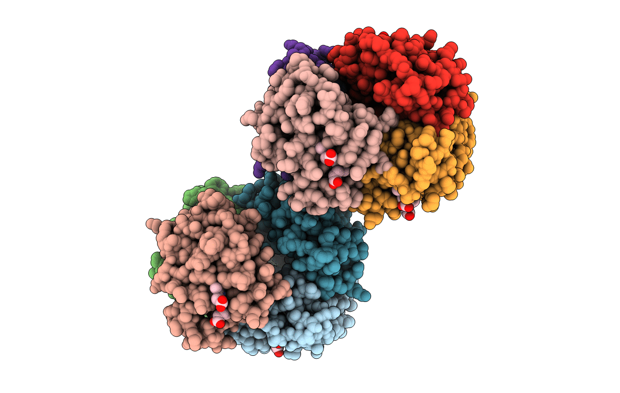
1HBS image
Entry Detail
PDB ID:
1HBS
Title:
REFINED CRYSTAL STRUCTURE OF DEOXYHEMOGLOBIN S. I. RESTRAINED LEAST-SQUARES REFINEMENT AT 3.0-ANGSTROMS RESOLUTION
Biological Source:
Source Organism:
PDB Version:
Deposition Date:
1982-06-02
Release Date:
1982-07-29
Method Details:
Experimental Method:
Resolution:
3.00 Å
R-Value Observed:
0.25
Space Group:
P 1 21 1
Macromolecular Entities
Polymer Type:polypeptide(L)
Description:HEMOGLOBIN S (DEOXY) (ALPHA CHAIN)
Chain IDs:A, C, E, G
Chain Length:141
Number of Molecules:4
Biological Source:Homo sapiens
Polymer Type:polypeptide(L)
Description:HEMOGLOBIN S (DEOXY) (BETA CHAIN)
Chain IDs:B, D, F, H
Chain Length:146
Number of Molecules:4
Biological Source:Homo sapiens
Ligand Molecules
Primary Citation
Refined crystal structure of deoxyhemoglobin S. I. Restrained least-squares refinement at 3.0-A resolution.
J.Biol.Chem. 260 8272 8279 (1985)
PMID: 4008491

Abstact

The crystal structure of deoxyhemoglobin S has been refined at 3.0-A resolution using the Hendrickson-Konnert restrained least-squares method. Comparison with the structure of deoxyhemoglobin A reveals a hingelike movement of the beta-chain A helices, which are involved in molecular contacts, toward the EF corners of their respective subunits. This movement brings the amino termini of the beta-chains closer to the molecular dyad. The A helices remain alpha-helical throughout their entire lengths. No other major structural difference is found between deoxyhemoglobin A and deoxyhemoglobin S.

Legend

Protein

Chemical

Disease

Primary Citation of related structures