Deposition Date
1993-02-05
Release Date
1994-01-31
Last Version Date
2024-11-06
Entry Detail
PDB ID:
1HBP
Keywords:
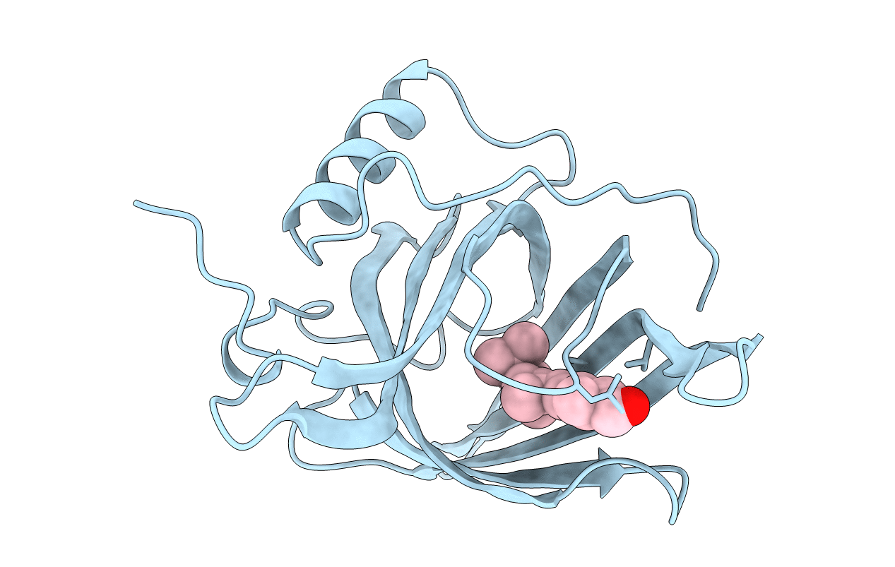
Title:
CRYSTAL STRUCTURE OF LIGANDED AND UNLIGANDED FORMS OF BOVINE PLASMA RETINOL-BINDING PROTEIN
Biological Source:
Source Organism(s):
Bos taurus (Taxon ID: 9913)
Method Details:
Experimental Method:
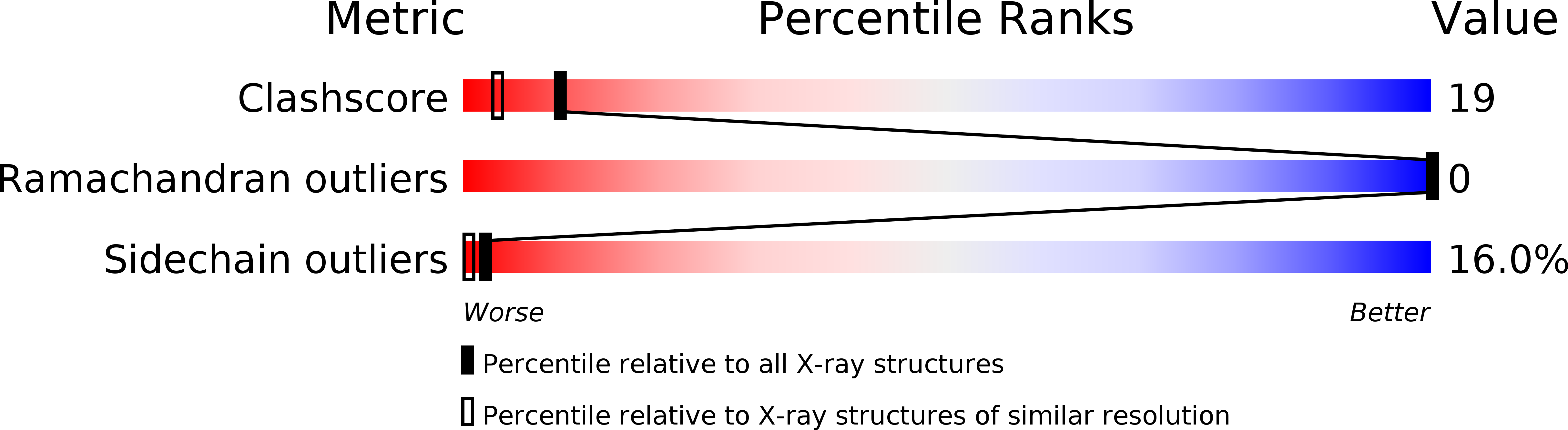
Resolution:
1.90 Å
R-Value Work:
0.19
R-Value Observed:
0.19
Space Group:
P 21 21 21