1HBI image
Deposition Date 1994-06-22
Release Date 1994-10-15
Last Version Date 2024-02-07
Entry Detail
PDB ID:
1HBI
Title:
CRYSTAL STRUCTURE OF OXYGENATED SCAPHARCA DIMERIC HEMOGLOBIN AT 1.7 ANGSTROMS RESOLUTION
Biological Source:
Source Organism(s):
Method Details:
Experimental Method:
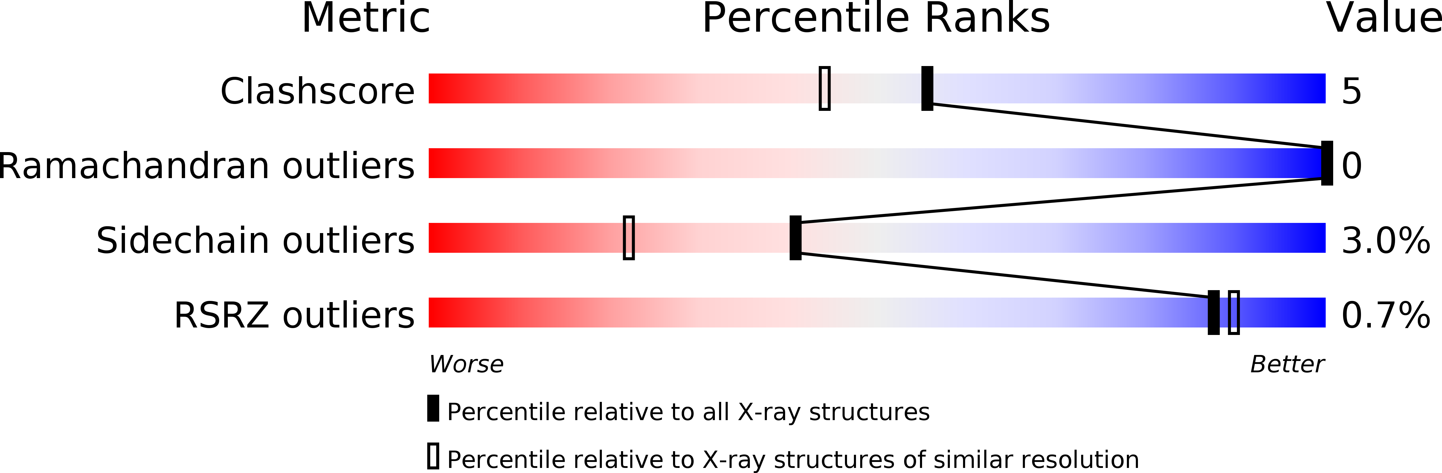
Resolution:
1.70 Å
R-Value Observed:
0.15
Space Group:
C 1 2 1
Macromolecular Entities
Structures with similar UniProt ID
Protein Blast
Polymer Type:polypeptide(L)
Molecule:HEMOGLOBIN (OXY)
Chain IDs:A, B
Chain Length:146
Number of Molecules:2
Biological Source:Scapharca inaequivalvis
Primary Citation
Crystal structure of oxygenated Scapharca dimeric hemoglobin at 1.7-A resolution.
J.Biol.Chem. 269 25259 25267 (1994)
PMID: 7929217

Abstact

The crystal structure of the cooperative dimeric hemoglobin from the blood clam Scapharca inaequivalvis has been determined in the oxygenated state and refined to an R-factor of 0.157 at 1.7-A resolution. The structure is very similar to the carbon monoxide-liganded form with subtle differences in ligand binding geometry. Oxygen binds to the heme iron in a bent conformation with Fe-O-O angles of 135 degrees and 150 degrees for the two subunits. These observed angles are lower than the equivalent angles in the carbon monoxide-liganded form and intermediate between the angles observed in structures of oxygenated sperm whale myoglobin and oxygenated human hemoglobin. This third high resolution structure of Scapharca dimeric hemoglobin permits a detailed analysis of the water structure in the highly hydrated interface between subunits.

Legend

Protein

Chemical

Disease

Primary Citation of related structures
Feedback Form
Name
Email
Institute
Feedback