Deposition Date
1992-01-07
Release Date
1994-01-31
Last Version Date
2024-05-22
Entry Detail
PDB ID:
1HBB
Keywords:
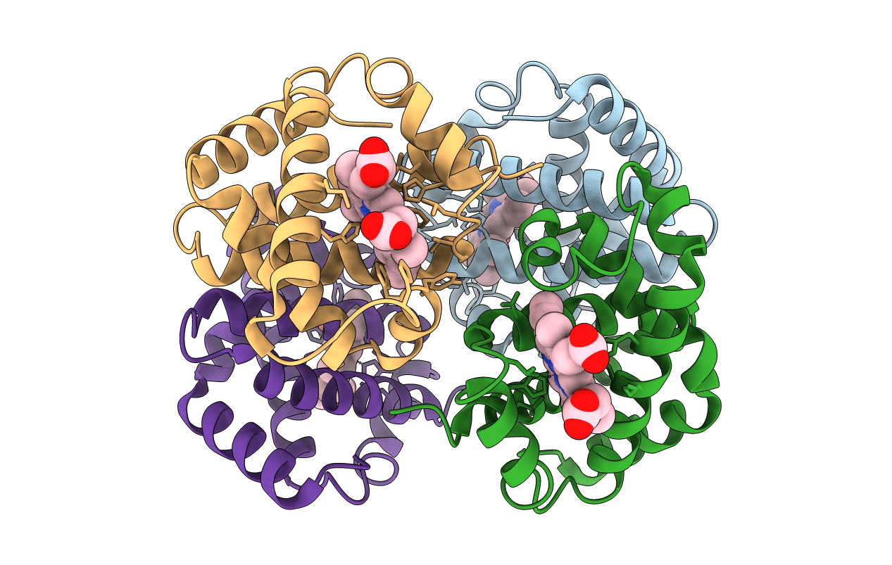
Title:
HIGH-RESOLUTION X-RAY STUDY OF DEOXYHEMOGLOBIN ROTHSCHILD 37BETA TRP-> ARG: A MUTATION THAT CREATES AN INTERSUBUNIT CHLORIDE-BINDING SITE
Biological Source:
Source Organism(s):
Homo sapiens (Taxon ID: 9606)
Method Details:
Experimental Method:
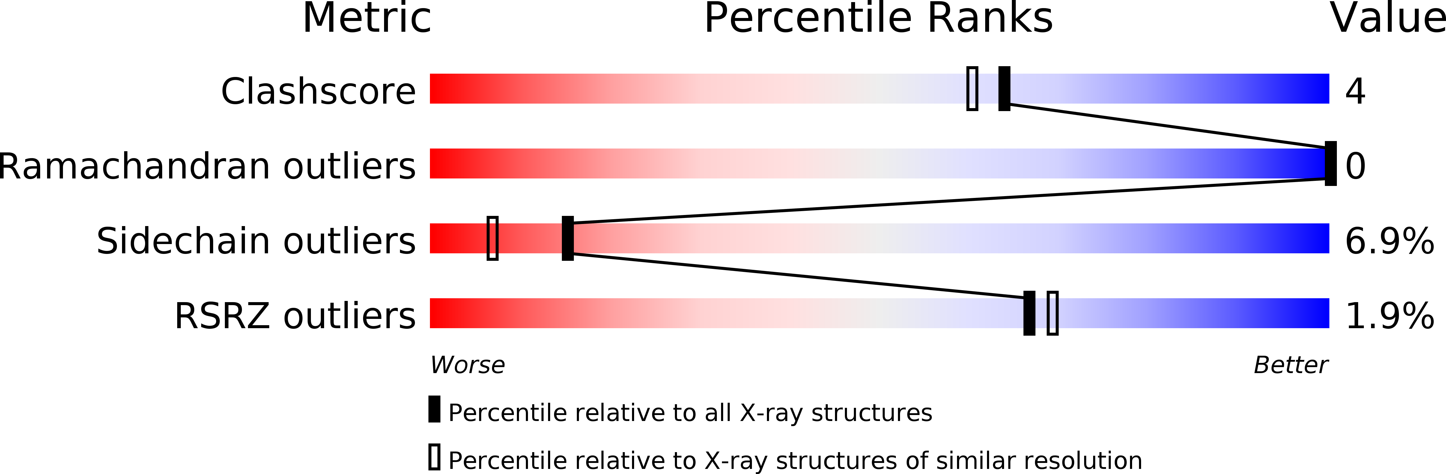
Resolution:
1.90 Å
R-Value Observed:
0.18
Space Group:
P 21 21 2