Deposition Date
2001-04-12
Release Date
2002-03-11
Last Version Date
2023-12-13
Entry Detail
PDB ID:
1HB6
Keywords:
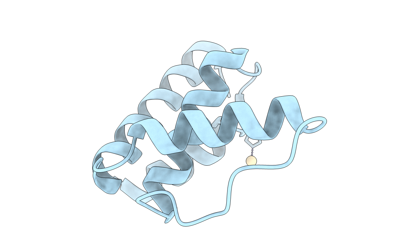
Title:
Structure of bovine Acyl-CoA binding protein in orthorhombic crystal form
Biological Source:
Source Organism(s):
BOS TAURUS (Taxon ID: 9913)
Method Details:
Experimental Method:
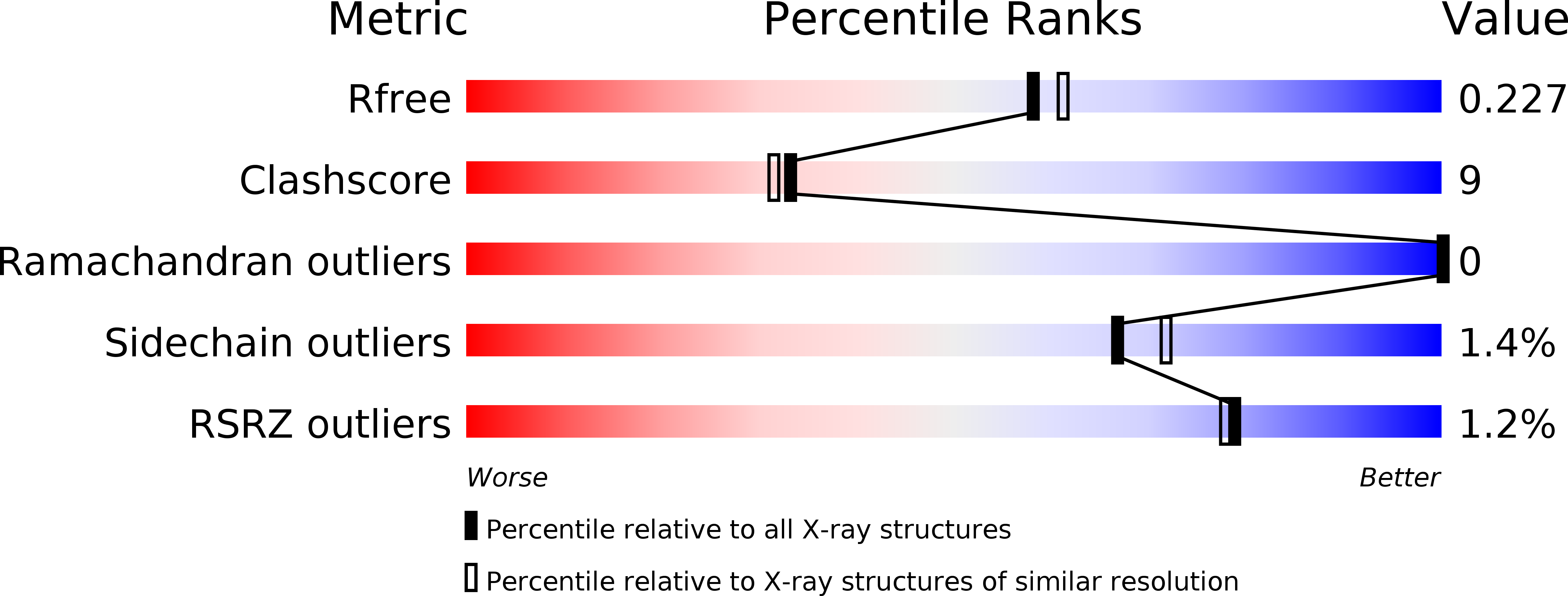
Resolution:
2.00 Å
R-Value Free:
0.22
R-Value Work:
0.20
R-Value Observed:
0.20
Space Group:
P 21 21 21