Deposition Date
2001-03-27
Release Date
2001-11-20
Last Version Date
2023-12-13
Entry Detail
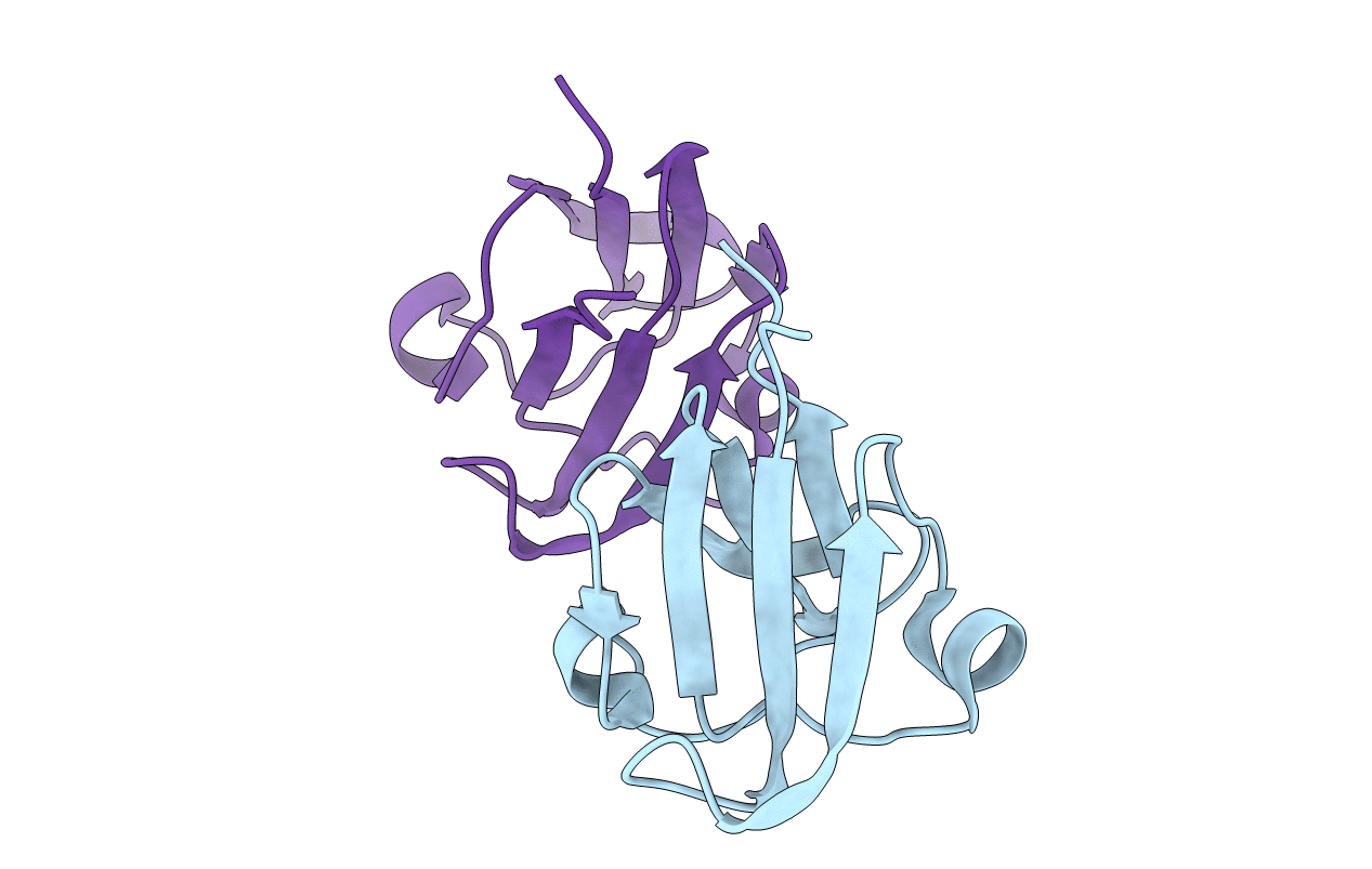
PDB ID:
1HA4
Keywords:
Title:
GammaS crystallin C terminal domain from Homo Sapiens
Biological Source:
Source Organism(s):
HOMO SAPIENS (Taxon ID: 9606)
Expression System(s):
Method Details:
Experimental Method:
Resolution:
2.40 Å
R-Value Free:
0.26
R-Value Work:
0.21
R-Value Observed:
0.21
Space Group:
P 65 2 2