1H9A image
Deposition Date 2001-03-06
Release Date 2001-05-03
Last Version Date 2023-12-13
Entry Detail
PDB ID:
1H9A
Title:
COMPLEX OF ACTIVE MUTANT (Q365->C) OF GLUCOSE 6-PHOSPHATE DEHYDROGENASE FROM L. MESENTEROIDES WITH COENZYME NADP
Biological Source:
Source Organism:
Host Organism:
Method Details:
Experimental Method:
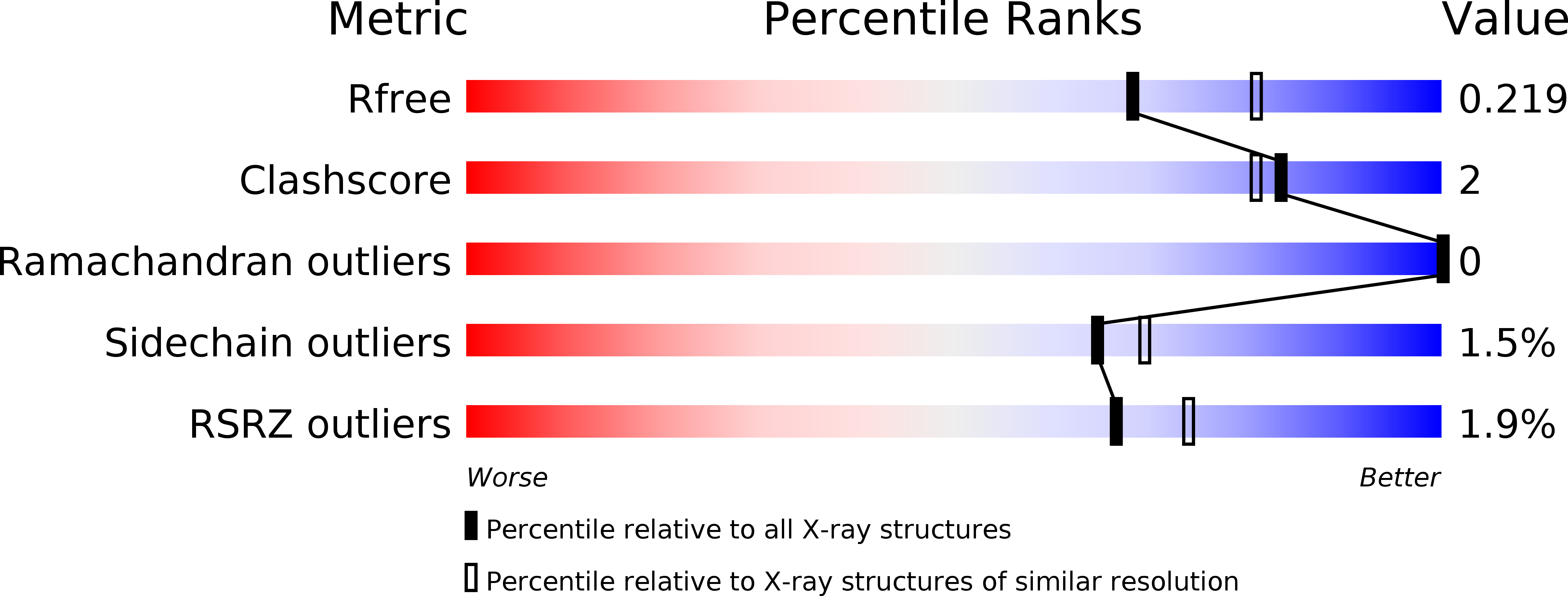
Resolution:
2.16 Å
R-Value Free:
0.22
R-Value Work:
0.18
R-Value Observed:
0.18
Space Group:
P 62 2 2
Macromolecular Entities
Structures with similar UniProt ID
Protein Blast
Polymer Type:polypeptide(L)
Molecule:GLUCOSE 6-PHOSPHATE 1-DEHYDROGENASE
Gene (Uniprot):zwf
Mutagens:YES
Chain IDs:A
Chain Length:485
Number of Molecules:1
Biological Source:LEUCONOSTOC MESENTEROIDES
Primary Citation
Nadp+ and Nad+ Binding to the Dual Coenzyme Specific Enzyme Leuconostoc Mesenteroides Glucose 6-Phosphate Dehydrogenase: Different Interdomain Hinge Angles are Seen in Different Binary and Ternary Complexes
Acta Crystallogr.,Sect.D 57 635 ? (2001)
PMID: 11320304 DOI: 10.1107/S0907444901003420

Abstact

The reduced coenzymes NADH and NADPH only differ by one phosphate, but in the cell NADH provides reducing power for catabolism while NADPH is utilized in biosynthetic pathways. Enzymes almost invariably discriminate between the coenzymes, but glucose 6-phosphate dehydrogenase (G6PD) from Leuconostoc mesenteroides is rare in being functionally dual specific. In order to elucidate the coenzyme selectivity, the structures of NADP(+)- and NAD(+)-complexed L. mesenteroides G6PD have been determined including data to 2.2 and 2.5 A resolution, respectively, and compared with unliganded G6PD crystallized in the same space groups. Coenzyme binding is also compared with that in a ternary complex of a mutant in which Asp177 in the active site has been mutated to asparagine. There are no gross structural differences between the complexes. In both binary complexes, the enzyme interdomain hinge angle has opened. NADP(+) binds to the furthest open form; of the residues within the coenzyme domain, only Arg46 moves, interacting with the 2'-phosphate and adenine. NAD(+) is less well defined in the binding site; smaller hinge opening is seen but larger local changes: Arg46 is displaced, Thr14 bonds the 3'-hydroxyl and Gln47 bonds the 2'-hydroxyl. In the ternary complex, the hinge angle has closed; only the adenine nucleotide is ordered in the binding site. Arg46 again provides most binding interactions.

Legend

Protein

Chemical

Disease

Primary Citation of related structures