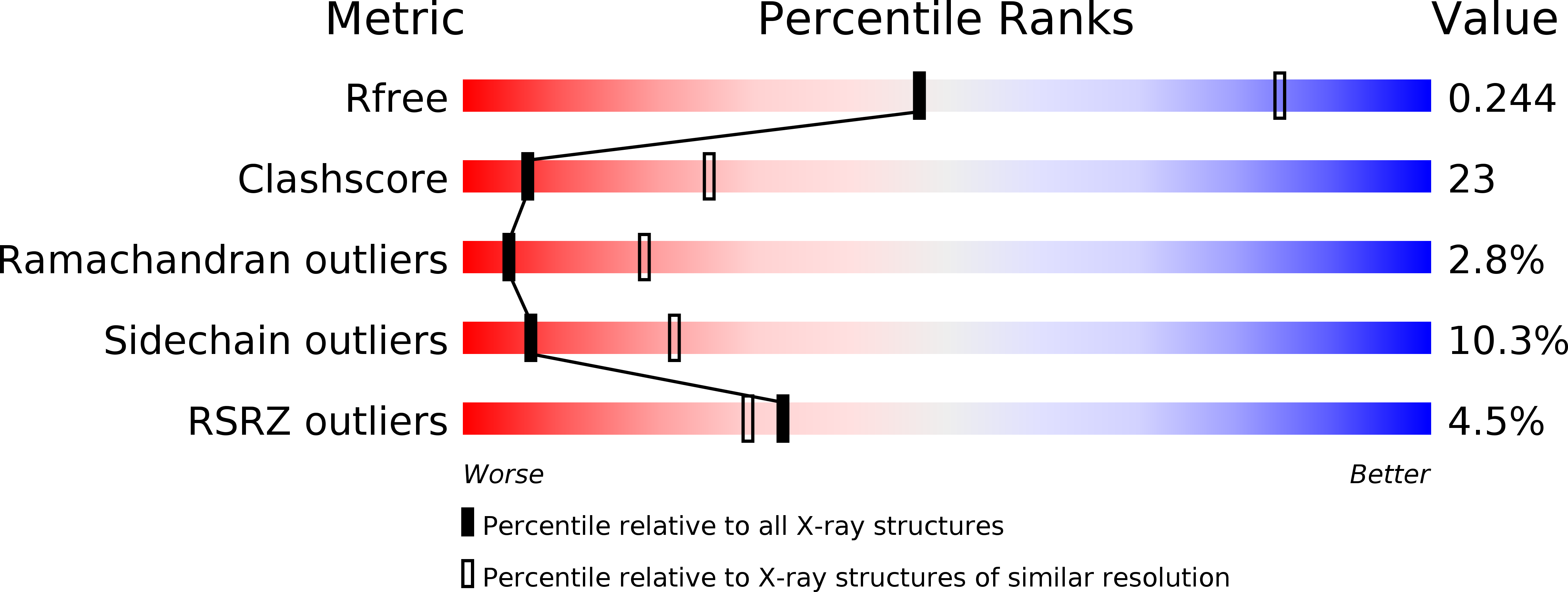
1H8T image
Deposition Date 2001-02-15
Release Date 2002-07-11
Last Version Date 2023-12-13
Entry Detail
PDB ID:
1H8T
Keywords:
Title:
Echovirus 11
Biological Source:
Source Organism:
ECHOVIRUS 11 (Taxon ID: 12078)
Method Details:
Experimental Method:
Resolution:
2.90 Å
R-Value Free:
0.25
R-Value Work:
0.24
R-Value Observed:
0.24
Space Group:
H 3 2
Macromolecular Entities
Polymer Type:polypeptide(L)
Molecule:ECHOVIRUS 11 COAT PROTEIN VP1
Chain IDs:A
Chain Length:292
Number of Molecules:1
Biological Source:ECHOVIRUS 11
Polymer Type:polypeptide(L)
Molecule:ECHOVIRUS 11 COAT PROTEIN VP2
Chain IDs:B
Chain Length:262
Number of Molecules:1
Biological Source:ECHOVIRUS 11
Polymer Type:polypeptide(L)
Molecule:ECHOVIRUS 11 COAT PROTEIN VP3
Chain IDs:C
Chain Length:238
Number of Molecules:1
Biological Source:ECHOVIRUS 11
Polymer Type:polypeptide(L)
Molecule:ECHOVIRUS 11 COAT PROTEIN VP4
Chain IDs:D
Chain Length:68
Number of Molecules:1
Biological Source:ECHOVIRUS 11
Primary Citation
Determination of the Structure of a Decay Accelerating Factor-Binding Clinical Isolate of Echovirus 11 Allows Mapping of Mutants with Altered Receptor Requirements for Infection
J.Virol. 76 7694 ? (2002)
PMID: 12097583 DOI: 10.1128/JVI.76.15.7694-7704.2002

Abstact

We have used X-ray crystallography to determine the structure of a decay accelerating factor (DAF)-binding, clinic-derived isolate of echovirus 11 (EV11-207). The structures of the capsid proteins closely resemble those of capsid proteins of other picornaviruses. The structure allows us to interpret a series of amino acid changes produced by passaging EV11-207 in different cell lines as highlighting the locations of multiple receptor-binding sites on the virion surface. We suggest that a DAF-binding site is located at the fivefold axes of the virion, while the binding site for a distinct but as yet unidentified receptor is located within the canyon surrounding the virion fivefold axes.

Legend

Protein

Chemical

Disease

Primary Citation of related structures