1H8N image
Deposition Date 2001-02-14
Release Date 2001-08-02
Last Version Date 2024-11-13
Entry Detail
PDB ID:
1H8N
Keywords:
Title:
Three-dimensional structure of anti-ampicillin single chain Fv fragment from phage-displayed murine antibody libraries
Biological Source:
Source Organism:
MUS MUSCULUS (Taxon ID: 10090)
Host Organism:
Method Details:
Experimental Method:
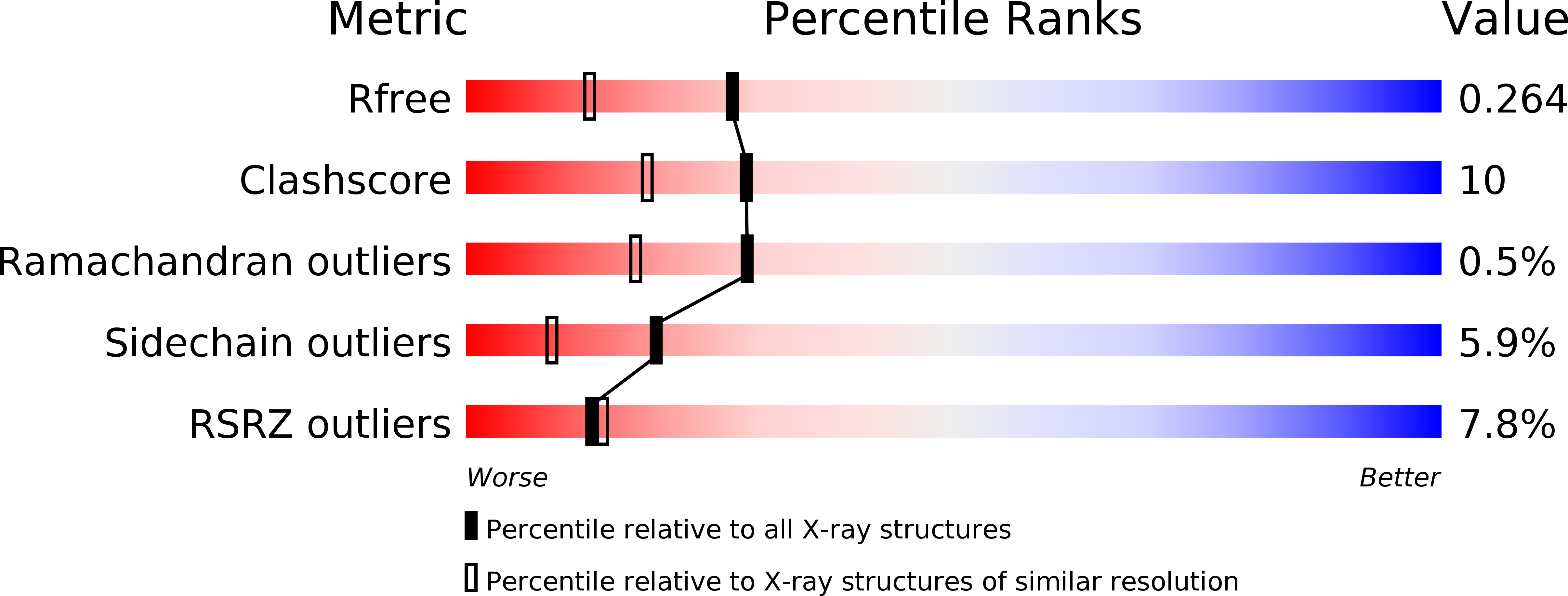
Resolution:
1.87 Å
R-Value Free:
0.27
R-Value Work:
0.23
R-Value Observed:
0.23
Space Group:
P 41 21 2
Macromolecular Entities
Polymer Type:polypeptide(L)
Molecule:MUTANT AL2 6E7S9G
Chain IDs:A
Chain Length:252
Number of Molecules:1
Biological Source:MUS MUSCULUS
Primary Citation
The Importance of Framework Residues H6, H7 and H10 in Antibody Heavy Chains: Experimental Evidence for a New Structural Subclassification of Antibody V(H) Domains
J.Mol.Biol. 309 701 ? (2001)
PMID: 11397090 DOI: 10.1006/JMBI.2001.4665

Abstact

The N-terminal segment (FR-H1) of the heavy chain (V(H)) of antibodies shows significant conformational variability correlating with the nature of the amino acids H6, H7 and H10 (Kabat H9). In this study, we have established a causal relationship between the local sequence and the structure of this framework region and linked this relationship to important biophysical properties such as affinity, folding yield and stability. We have generated six mutants of the scFv fragment aL2, covering some of the most abundant amino acid combinations in positions H6, H7 and H10 (according to a new consensus nomenclature, Kabat H9). For the aL2 wild-type (w.t.) with the sequence 6(Q)7(P)10(A) and for two of the mutants, the X-ray structures have been determined. The structure of the triple mutant aL2-6(E)7(S)10(G) shows the FR-H1 backbone conformations predicted for this amino acid combination, which is distinctly different from the structure of the w.t, thus supporting our hypothesis that these residues determine the conformation of this segment. The mutant aL2-6(E)7(P)10(G) represents a residue combination not occurring in natural antibody sequences. It shows a completely different, unique structure in the first beta-strand of V(H), not observed in natural Fv fragments and forms a novel type of diabody. Two V(H) domains of the mutant associate by swapping the first beta-strand. Concentration-dependent changes in Trp fluorescence indicate that this dimerization also occurs in solution. The mutations in amino acids H6, H7 and H10 (Kabat H9) influence the dimerization behavior of the scFv and its thermodynamic stability. All the observations reported here have practical implications for the cloning of Fv fragments with degenerate primers, as well as for the design of new antibodies by CDR grafting or synthetic libraries.

Legend

Protein

Chemical

Disease

Primary Citation of related structures