
Deposition Date
2001-01-22
Release Date
2001-11-27
Last Version Date
2024-11-20
Entry Detail
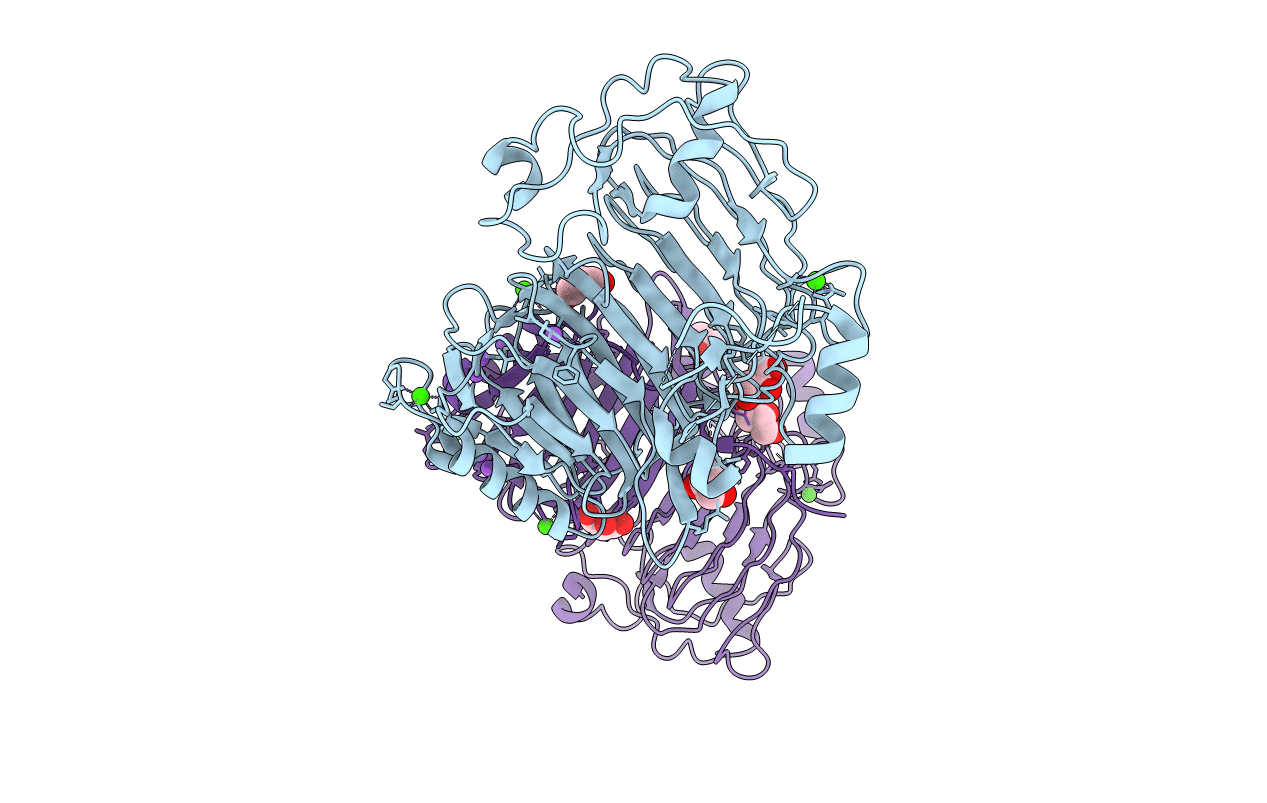
PDB ID:
1H80
Keywords:
Title:
1,3-ALPHA-1,4-BETA-D-GALACTOSE-4-SULFATE- 3,6-ANHYDRO-D-GALACTOSE-2-SULFATE 4 GALACTOHYDROLASE
Biological Source:
Source Organism(s):
ALTEROMONAS SP. ATCC43554 (Taxon ID: 116059)
Expression System(s):
Method Details:
Experimental Method:
Resolution:
1.60 Å
R-Value Free:
0.22
R-Value Work:
0.20
R-Value Observed:
0.20
Space Group:
P 1 21 1
