
Deposition Date
2001-01-19
Release Date
2001-02-23
Last Version Date
2024-05-01
Entry Detail
PDB ID:
1H7X
Keywords:
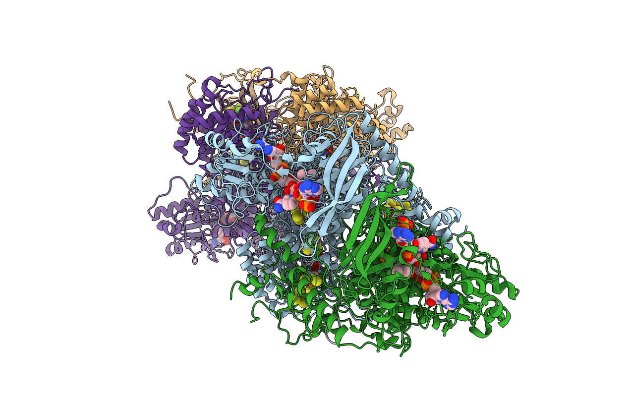
Title:
Dihydropyrimidine dehydrogenase (DPD) from pig, ternary complex of a mutant enzyme (C671A), NADPH and 5-fluorouracil
Biological Source:
Source Organism(s):
SUS SCROFA (Taxon ID: 9823)
Expression System(s):
Method Details:
Experimental Method:
Resolution:
2.01 Å
R-Value Free:
0.19
R-Value Work:
0.17
R-Value Observed:
0.17
Space Group:
P 1 21 1
