1H6H image
Deposition Date 2001-06-15
Release Date 2001-11-01
Last Version Date 2024-05-08
Entry Detail
PDB ID:
1H6H
Keywords:
Title:
Structure of the PX domain from p40phox bound to phosphatidylinositol 3-phosphate
Biological Source:
Source Organism:
HOMO SAPIENS (Taxon ID: 9606)
Method Details:
Experimental Method:
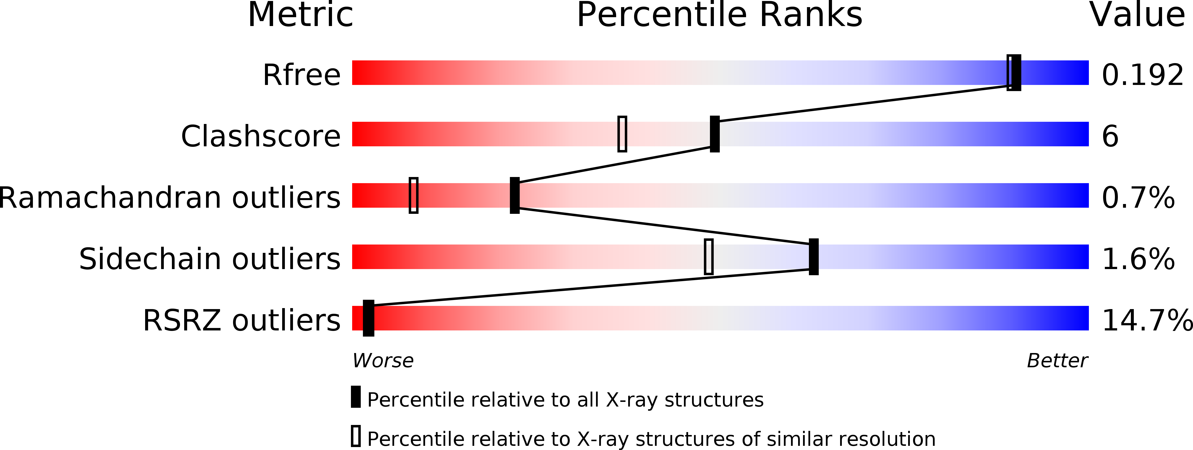
Resolution:
1.70 Å
R-Value Free:
0.23
R-Value Work:
0.19
R-Value Observed:
0.19
Space Group:
C 1 2 1
Macromolecular Entities
Polymer Type:polypeptide(L)
Molecule:NEUTROPHIL CYTOSOL FACTOR 4
Gene (Uniprot):NCF4
Chain IDs:A
Chain Length:143
Number of Molecules:1
Biological Source:HOMO SAPIENS
Primary Citation
The Crystal Structure of the Px Domain from P40Phox Bound to Phosphatidylinositol 3-Phosphate
Mol.Cell 8 829 ? (2001)
PMID: 11684018 DOI: 10.1016/S1097-2765(01)00372-0

Abstact

More than 50 human proteins with a wide range of functions have a 120 residue phosphoinositide binding module known as the PX domain. The 1.7 A X-ray crystal structure of the PX domain from the p40(phox) subunit of NADPH oxidase bound to PtdIns(3)P shows that the PX domain embraces the 3-phosphate on one side of a water-filled, positively charged pocket and reveals how 3-phosphoinositide specificity is achieved. A chronic granulomatous disease (CGD)-associated mutation in the p47(phox) PX domain that abrogates PtdIns(3)P binding maps to a conserved Arg that does not directly interact with the phosphoinositide but instead appears to stabilize a critical lipid binding loop. The SH3 domain present in the full-length protein does not affect soluble PtdIns(3)P binding to the p40(phox) PX domain.

Legend

Protein

Chemical

Disease

Primary Citation of related structures