1H6E image
Deposition Date 2001-06-12
Release Date 2001-11-28
Last Version Date 2023-12-13
Entry Detail
PDB ID:
1H6E
Title:
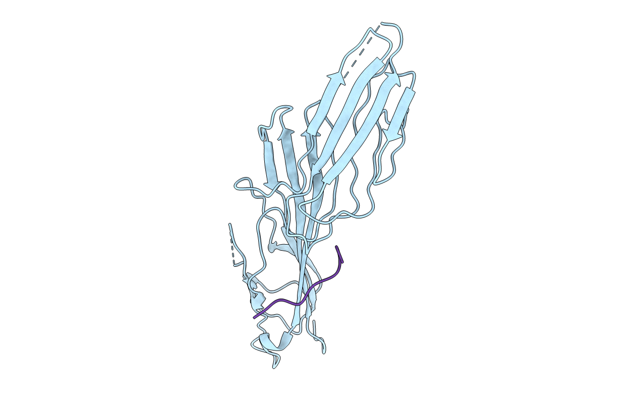
MU2 ADAPTIN SUBUNIT (AP50) OF AP2 ADAPTOR (SECOND DOMAIN), COMPLEXED WITH CTLA-4 INTERNALIZATION PEPTIDE TTGVYVKMPPT
Biological Source:
Source Organism:
HOMO SAPIENS (Taxon ID: 9606)
Host Organism:
Method Details:
Experimental Method:
Resolution:
3.60 Å
R-Value Free:
0.31
R-Value Work:
0.27
Space Group:
P 64
Macromolecular Entities
Polymer Type:polypeptide(L)
Molecule:CLATHRIN COAT ASSEMBLY PROTEIN AP50
Chain IDs:A
Chain Length:288
Number of Molecules:1
Biological Source:HOMO SAPIENS
Polymer Type:polypeptide(L)
Molecule:CYTOTOXIC T-LYMPHOCYTE PROTEIN 4
Gene (Uniprot):CTLA4
Chain IDs:B (auth: P)
Chain Length:11
Number of Molecules:1
Biological Source:HOMO SAPIENS
Ligand Molecules
Primary Citation
Study of the Interaction of the Medium Chain Mu2 Subunit of the Clathrin-Associated Adapter Protein Complex 2 with Cytotoxic T-Lymphocyte Antigen 4 and Cd28
Biochem.J. 359 427 ? (2001)
PMID: 11583591 DOI: 10.1042/0264-6021:3590427

Abstact

The medium chain mu 2 subunit (AP50) of the clathrin-associated adapter protein complex 2 (AP-2) interacts specifically with the tyrosine-based signals of several integral membrane proteins through the consensus sequence YXXPhi, where X can be any residue and Phi is a large hydrophobic residue. Using surface plasmon resonance combined with structural information, we have analysed the interaction of AP50 with peptides derived from the cytoplasmic tail of cytotoxic T-lymphocyte antigen 4 (CTLA-4). The crystal structure of AP50 in complex with a CTLA-4-derived peptide was determined to 3.6 A (1 A=0.1 nm) resolution. The binding domain of AP50 (residues 164-435) was expressed in Escherichia coli and purified. In agreement with previous reports, the AP50 domain bound to residues 152-174 of CTLA-4, but not to the same peptide that was phosphorylated at the single tyrosine residue (position 165). The interaction exhibited fast kinetics with rapid on and off rates and a K(d) of 0.7 microM. In order to further understand why AP50 binds to CTLA-4, but not to the homologous receptor CD28, a comparison of binding of AP50 with five peptides with single changes in and around the YXXPhi motif to the equivalent residues of CD28 was made. T162H greatly reduced binding, whereas T161L had little effect. Mutations G163S, V164D and K167N all exhibited reduced binding. Modelling of the single amino acid changes using structural information, was in broad agreement with the binding data, demonstrating that residues outside of the YXXPhi motif are also important in the interaction of membrane proteins with AP50.

Legend

Protein

Chemical

Disease

Primary Citation of related structures