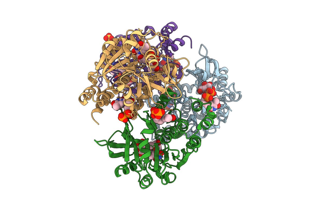
1H5T image
Deposition Date 2001-05-25
Release Date 2001-11-23
Last Version Date 2025-04-09
Entry Detail
PDB ID:
1H5T
Keywords:
Title:
Thymidylyltransferase complexed with Thymidylyldiphosphate-glucose
Biological Source:
Source Organism:
Escherichia coli (Taxon ID: 562)
Host Organism:
Method Details:
Experimental Method:
Resolution:
1.90 Å
R-Value Free:
0.22
R-Value Work:
0.17
Space Group:
P 1 21 1
Macromolecular Entities
Polymer Type:polypeptide(L)
Molecule:Glucose-1-phosphate thymidylyltransferase 1
Gene (Uniprot):rfbA
Chain IDs:A, C, D
Chain Length:293
Number of Molecules:3
Biological Source:Escherichia coli
Polymer Type:polypeptide(L)
Molecule:Glucose-1-phosphate thymidylyltransferase 1
Gene (Uniprot):rfbA
Chain IDs:B
Chain Length:293
Number of Molecules:1
Biological Source:Escherichia coli
Primary Citation
Kinetic and Crystallographic Analyses Support a Sequential-Ordered Bi Bi Catalytic Mechanism for Escherichia Coli Glucose-1-Phosphate Thymidylyltransferase
J.Mol.Biol. 313 831 ? (2001)
PMID: 11697907 DOI: 10.1006/JMBI.2001.5073

Abstact

Glucose-1-phosphate thymidylyltransferase is the first enzyme in the biosynthesis of dTDP-l-rhamnose, the precursor of l-rhamnose, an essential component of surface antigens, such as the O-lipopolysaccharide, mediating virulence and adhesion to host tissues in many microorganisms. The enzyme catalyses the formation of dTDP-glucose, from dTTP and glucose 1-phosphate, as well as its pyrophosphorolysis. To shed more light on the catalytic properties of glucose-1-phosphate thymidylyltransferase from Escherichia coli, specifically distinguishing between ping pong and sequential ordered bi bi reaction mechanisms, the enzyme kinetic properties have been analysed in the presence of different substrates and inhibitors. Moreover, three different complexes of glucose-1-phosphate thymidylyltransferase (co-crystallized with dTDP, with dTMP and glucose-1-phosphate, with d-thymidine and glucose-1-phosphate) have been analysed by X-ray crystallography, in the 1.9-2.3 A resolution range (R-factors of 17.3-17.5 %). The homotetrameric enzyme shows strongly conserved substrate/inhibitor binding modes in a surface cavity next to the topological switch-point of a quasi-Rossmann fold. Inspection of the subunit tertiary structure reveals relationships to other enzymes involved in the biosynthesis of nucleotide-sugars, including distant proteins such as the molybdenum cofactor biosynthesis protein MobA. The precise location of the substrate relative to putative reactive residues in the catalytic center suggests that, in keeping with the results of the kinetic measurements, both catalysed reactions, i.e. dTDP-glucose biosynthesis and pyrophosphorolysis, follow a sequential ordered bi bi catalytic mechanism.

Legend

Protein

Chemical

Disease

Primary Citation of related structures