Deposition Date
2001-05-16
Release Date
2001-07-06
Last Version Date
2023-12-13
Entry Detail
PDB ID:
1H4Z
Keywords:
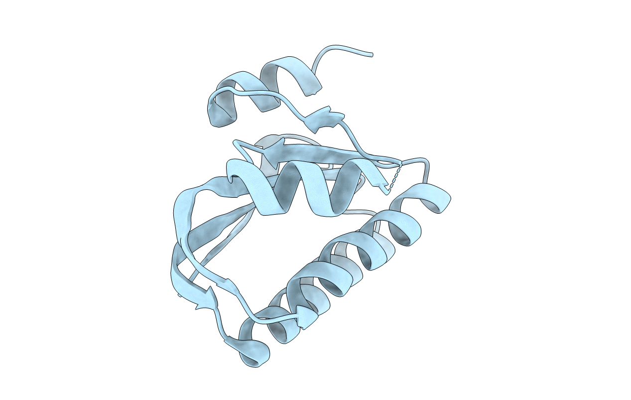
Title:
Structure of the Anti-Sigma Factor Antagonist SpoIIAA in its Unphosphorylated Form
Biological Source:
Source Organism(s):
BACILLUS SPHAERICUS (Taxon ID: 1421)
Expression System(s):
Method Details:
Experimental Method:
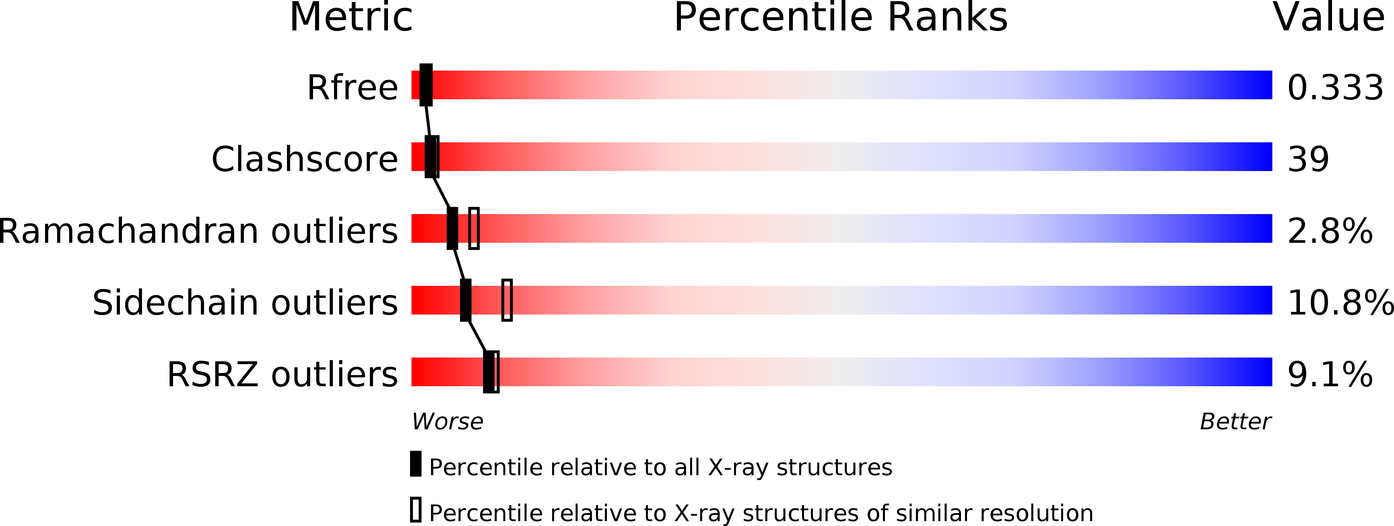
Resolution:
2.74 Å
R-Value Free:
0.30
R-Value Work:
0.25
Space Group:
P 65 2 2