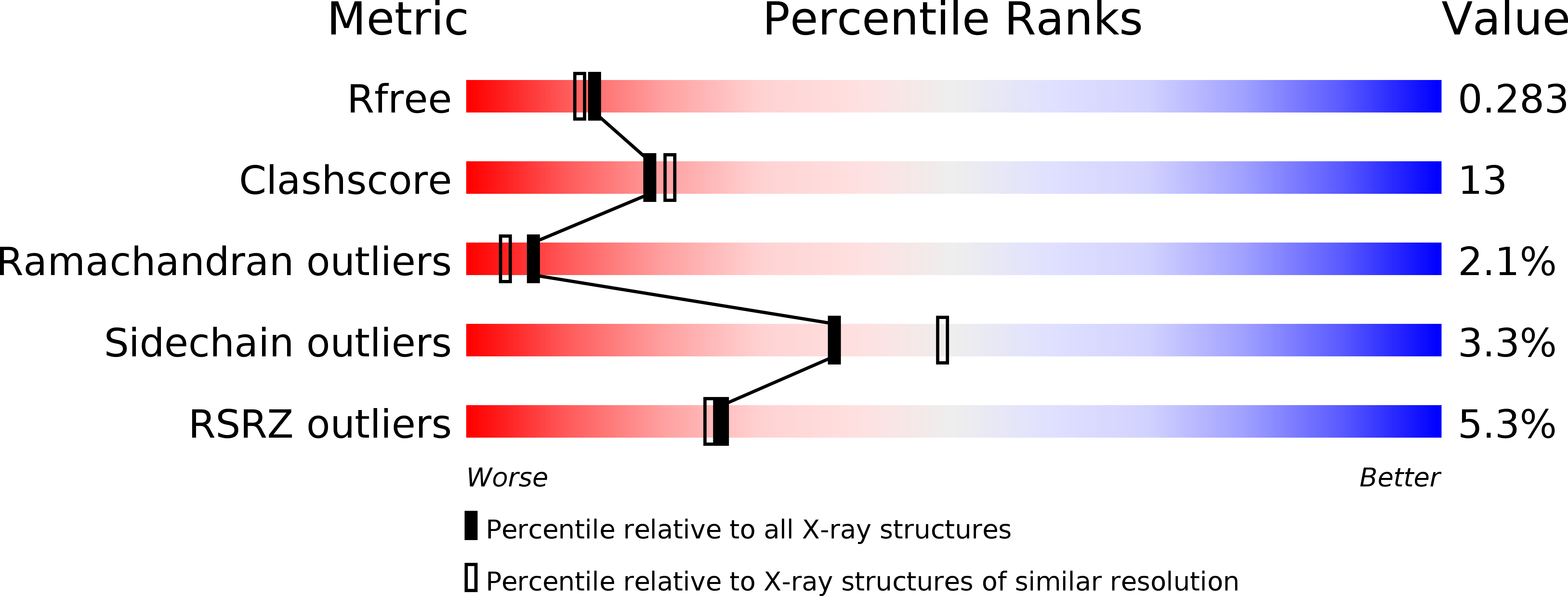
1H4U image
Deposition Date 2001-05-14
Release Date 2001-06-28
Last Version Date 2024-11-06
Entry Detail
PDB ID:
1H4U
Title:
Domain G2 of mouse nidogen-1
Biological Source:
Source Organism:
MUS MUSCULUS (Taxon ID: 10090)
Host Organism:
Method Details:
Experimental Method:
Resolution:
2.20 Å
R-Value Free:
0.28
R-Value Work:
0.24
R-Value Observed:
0.24
Space Group:
P 64
Macromolecular Entities
Structures with similar UniProt ID
Protein Blast
Polymer Type:polypeptide(L)
Molecule:NIDOGEN-1
Gene (Uniprot):Nid1
Chain IDs:A
Chain Length:265
Number of Molecules:1
Biological Source:MUS MUSCULUS
Primary Citation
Crystal Structure and Mutational Analysis of a Perlecan-Binding Fragment of Nidogen-1
Nat.Struct.Biol. 8 634 ? (2001)
PMID: 11427896 DOI: 10.1038/89683

Abstact

Nidogen, an invariant component of basement membranes, is a multifunctional protein that interacts with most other major basement membrane proteins. Here, we report the crystal structure of the mouse nidogen-1 G2 fragment, which contains binding sites for collagen IV and perlecan. The structure is composed of an EGF-like domain and an 11-stranded beta-barrel with a central helix. The beta-barrel domain has unexpected similarity to green fluorescent protein. A large surface patch on the beta-barrel is strikingly conserved in all metazoan nidogens. Site-directed mutagenesis demonstrates that the conserved residues are involved in perlecan binding.

Legend

Protein

Chemical

Disease

Primary Citation of related structures