1H4P image
Deposition Date 2001-05-11
Release Date 2003-10-02
Last Version Date 2024-11-13
Entry Detail
PDB ID:
1H4P
Keywords:
Title:
Crystal structure of exo-1,3-beta glucanse from Saccharomyces cerevisiae
Biological Source:
Source Organism:
Method Details:
Experimental Method:
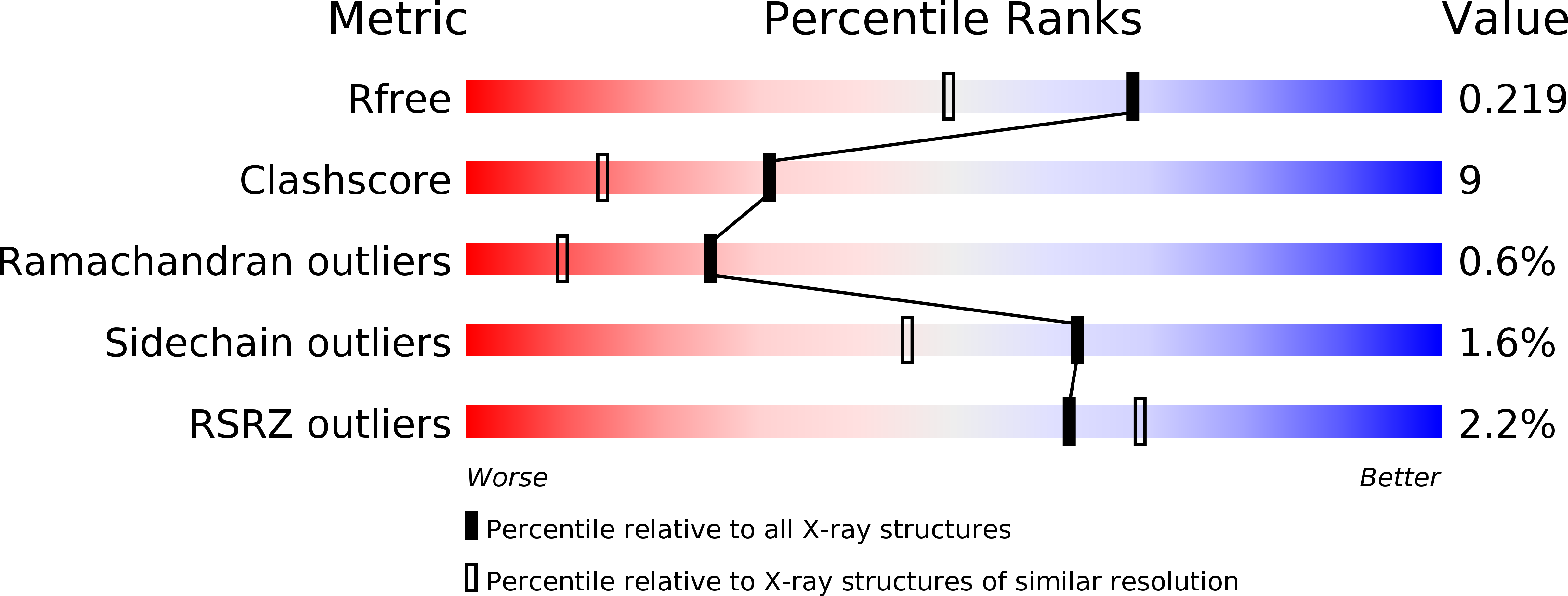
Resolution:
1.75 Å
R-Value Free:
0.22
R-Value Work:
0.20
R-Value Observed:
0.20
Space Group:
P 41 21 2
Macromolecular Entities
Polymer Type:polypeptide(L)
Molecule:GLUCAN 1,3-BETA-GLUCOSIDASE I/II
Gene (Uniprot):EXG1
Chain IDs:A, B
Chain Length:408
Number of Molecules:2
Biological Source:SACCHAROMYCES CEREVISIAE
Modified Residue
Compound ID Chain ID Parent Comp ID Details 2D Image
ASN A ASN GLYCOSYLATION SITE
Ligand Molecules
Primary Citation
The Er Protein Folding Sensor Udp-Glucose Glycoprotein:Glucosyltransferase Modifies Substrates Distant to Local Changes in Glycoprotein Conformation.
Nat.Struct.Mol.Biol. 11 128 ? (2004)
PMID: 14730348 DOI: 10.1038/NSMB715

Abstact

We present in vitro data that explain the recognition mechanism of misfolded glycoproteins by UDP-glucose glycoprotein-glucosyltransferase (UGGT). The glycoprotein exo-(1,3)-beta-glucanase (beta-Glc) bearing two glycans unfolds in a pH-dependent manner to become a misfolded substrate for UGGT. In the crystal structure of this glycoprotein, the local hydrophobicity surrounding each glycosylation site coincides with the differential recognition of N-linked glycans by UGGT. We introduced a single F280S point mutation, producing a beta-Glc protein with full enzymatic activity that was both recognized as misfolded and monoglucosylated by UGGT. Contrary to current views, these data show that UGGT can modify N-linked glycans positioned at least 40 A from localized regions of disorder and sense subtle conformational changes within structurally compact, enzymatically active glycoprotein substrates.

Legend

Protein

Chemical

Disease

Primary Citation of related structures