1H4L image
Deposition Date 2001-05-11
Release Date 2002-08-14
Last Version Date 2023-12-13
Entry Detail
PDB ID:
1H4L
Title:
Structure and regulation of the CDK5-p25(nck5a) complex
Biological Source:
Source Organism:
HOMO SAPIENS (Taxon ID: 9606)
Host Organism:
Method Details:
Experimental Method:
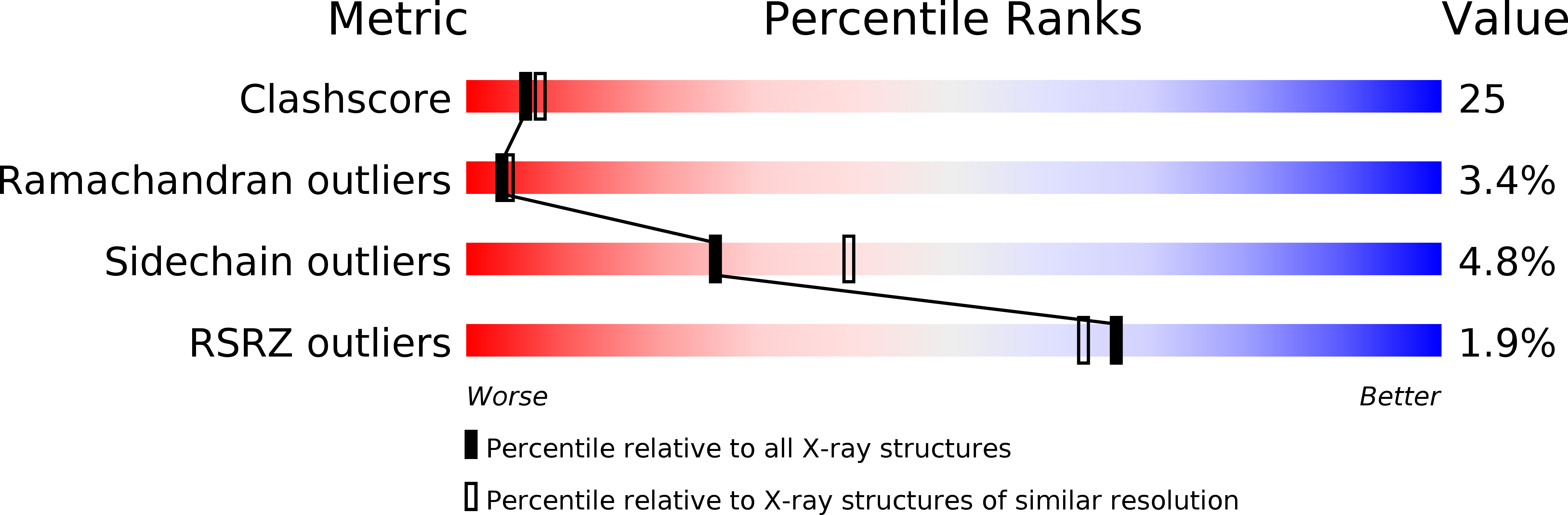
Resolution:
2.65 Å
R-Value Free:
0.28
R-Value Work:
0.23
R-Value Observed:
0.23
Space Group:
C 1 2 1
Macromolecular Entities
Structures with similar UniProt ID
Protein Blast
Polymer Type:polypeptide(L)
Molecule:CELL DIVISION PROTEIN KINASE 5
Gene (Uniprot):CDK5
Chain IDs:A, B
Chain Length:292
Number of Molecules:2
Biological Source:HOMO SAPIENS
Structures with similar UniProt ID
Protein Blast
Polymer Type:polypeptide(L)
Molecule:CYCLIN-DEPENDENT KINASE 5 ACTIVATOR
Gene (Uniprot):CDK5R1
Chain IDs:C (auth: D), D (auth: E)
Chain Length:147
Number of Molecules:2
Biological Source:HOMO SAPIENS
Primary Citation
Structure and Regulation of the Cdk5-P25(Nck5A) Complex
Mol.Cell 8 657 ? (2001)
PMID: 11583627 DOI: 10.1016/S1097-2765(01)00343-4

Abstact

CDK5 plays an indispensable role in the central nervous system, and its deregulation is involved in neurodegeneration. We report the crystal structure of a complex between CDK5 and p25, a fragment of the p35 activator. Despite its partial structural similarity with the cyclins, p25 displays an unprecedented mechanism for the regulation of a cyclin-dependent kinase. p25 tethers the unphosphorylated T loop of CDK5 in the active conformation. Residue Ser159, equivalent to Thr160 on CDK2, contributes to the specificity of the CDK5-p35 interaction. Its substitution with threonine prevents p35 binding, while the presence of alanine affects neither binding nor kinase activity. Finally, we provide evidence that the CDK5-p25 complex employs a distinct mechanism from the phospho-CDK2-cyclin A complex to establish substrate specificity.

Legend

Protein

Chemical

Disease

Primary Citation of related structures