1H3M image
Deposition Date 2002-09-10
Release Date 2003-08-01
Last Version Date 2024-11-13
Entry Detail
PDB ID:
1H3M
Keywords:
Title:
Structure of 4-diphosphocytidyl-2C-methyl-D-erythritol synthetase
Biological Source:
Source Organism:
Host Organism:
Method Details:
Experimental Method:
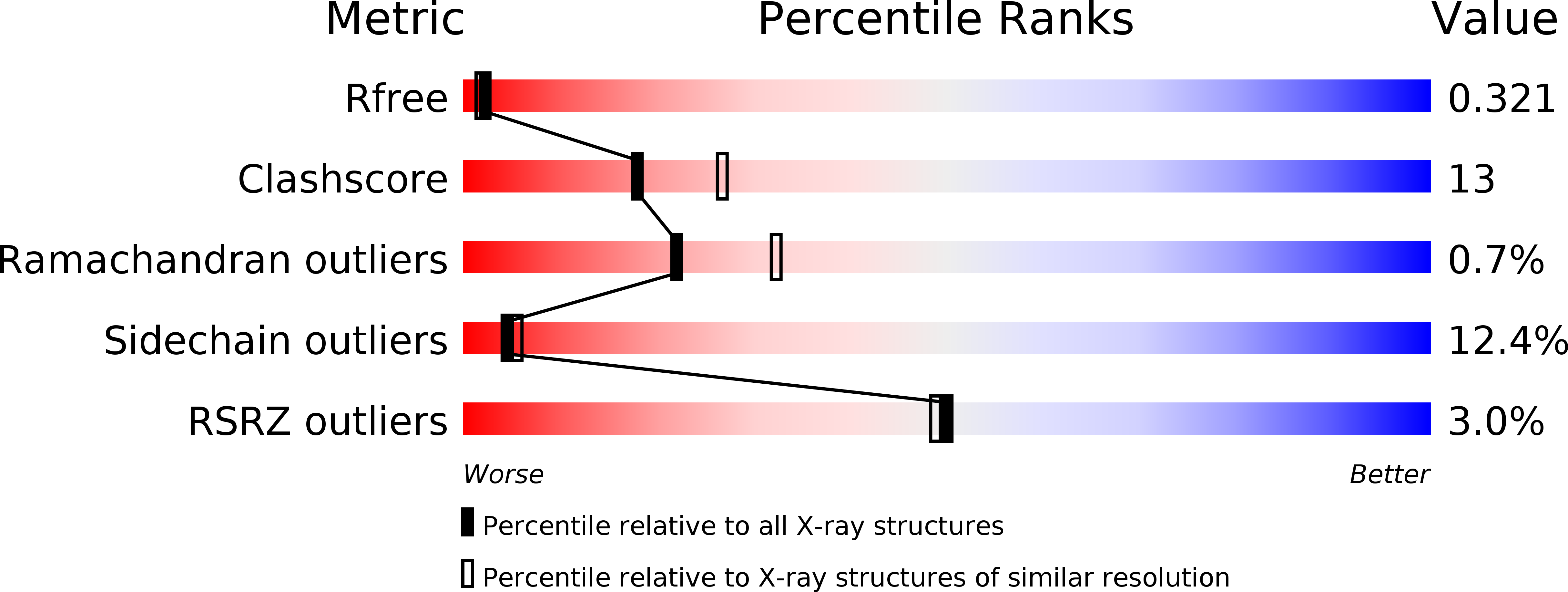
Resolution:
2.40 Å
R-Value Free:
0.33
R-Value Work:
0.23
R-Value Observed:
0.24
Space Group:
P 41 21 2
Macromolecular Entities
Structures with similar UniProt ID
Protein Blast
Polymer Type:polypeptide(L)
Molecule:2-C-METHYL-D-ERYTHRITOL 4-PHOSPHATE CYTIDYLYLTRANSFERASE
Gene (Uniprot):ispD
Mutagens:YES
Chain IDs:A, B
Chain Length:235
Number of Molecules:2
Biological Source:ESCHERICHIA COLI
Primary Citation
Structure of a Tetragonal Crystal Form of Escherichia Coli 2-C-Methyl-D-Erythritol 4-Phosphate Cytidylyltransferase
Acta Crystallogr.,Sect.D 59 607 ? (2003)
PMID: 12595740 DOI: 10.1107/S090744490202365X

Abstact

2-C-Methyl-D-erythritol 4-phosphate cytidylyltransferase is an essential enzyme in the mevalonate-independent pathway of isoprenoid biosynthesis. The structure of a tetragonal crystal form has been solved by molecular replacement and refined to 2.4 A resolution. Structure and sequence comparisons suggest that the enzyme is a suitable target for a structure-based approach to the development of novel broad-spectrum antibiotics. However, the absence of ligands in the enzyme active site together with the moderate resolution of the structure indicates that this tetragonal crystal form is inferior to that of a previously reported highly ordered monoclinic form [Richard et al. (2001), Nature Struct. Biol. 8, 641-647].

Legend

Protein

Chemical

Disease

Primary Citation of related structures
Feedback Form
Name
Email
Institute
Feedback