
Deposition Date
2002-08-28
Release Date
2002-09-12
Last Version Date
2024-05-01
Entry Detail
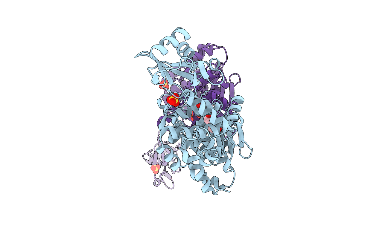
PDB ID:
1H3F
Keywords:
Title:
Tyrosyl-tRNA synthetase from Thermus thermophilus complexed with tyrosinol
Biological Source:
Source Organism(s):
THERMUS THERMOPHILUS (Taxon ID: 262724)
Expression System(s):
Method Details:
Experimental Method:
Resolution:
2.00 Å
R-Value Free:
0.26
R-Value Work:
0.23
R-Value Observed:
0.23
Space Group:
P 21 21 21
