1H2G image
Deposition Date 2002-08-08
Release Date 2003-07-17
Last Version Date 2023-12-13
Entry Detail
PDB ID:
1H2G
Keywords:
Title:
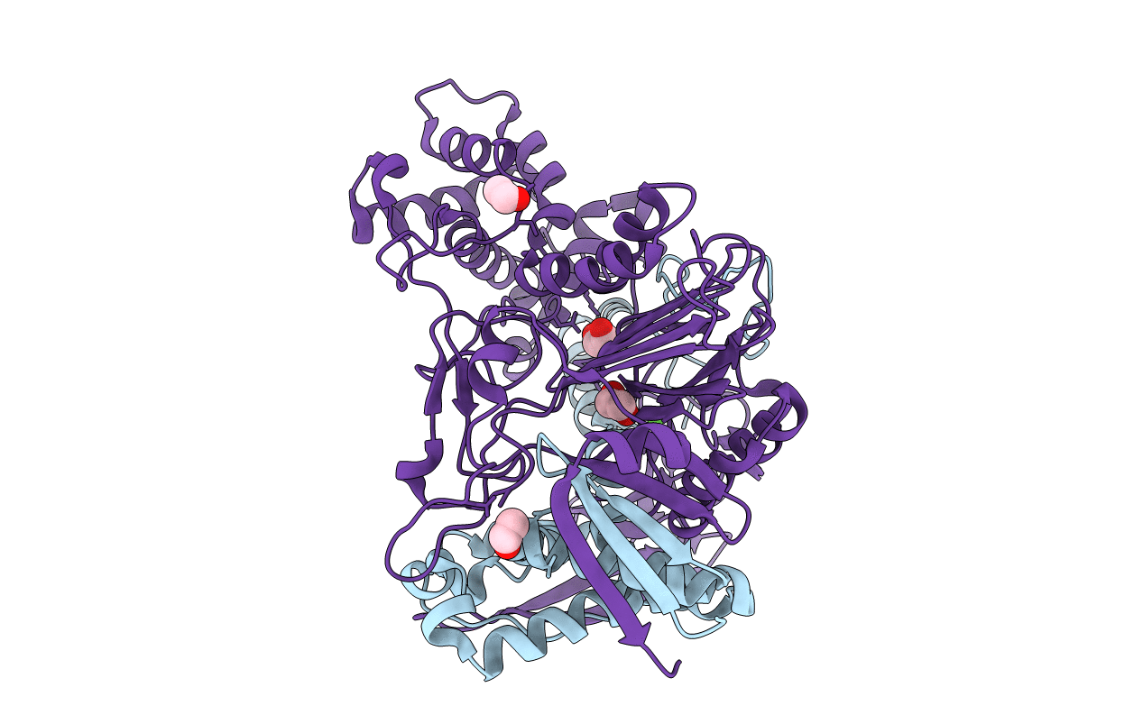
Altered substrate specificity mutant of penicillin acylase
Biological Source:
Source Organism:
ESCHERICHIA COLI (Taxon ID: 562)
Host Organism:
Method Details:
Experimental Method:
Resolution:
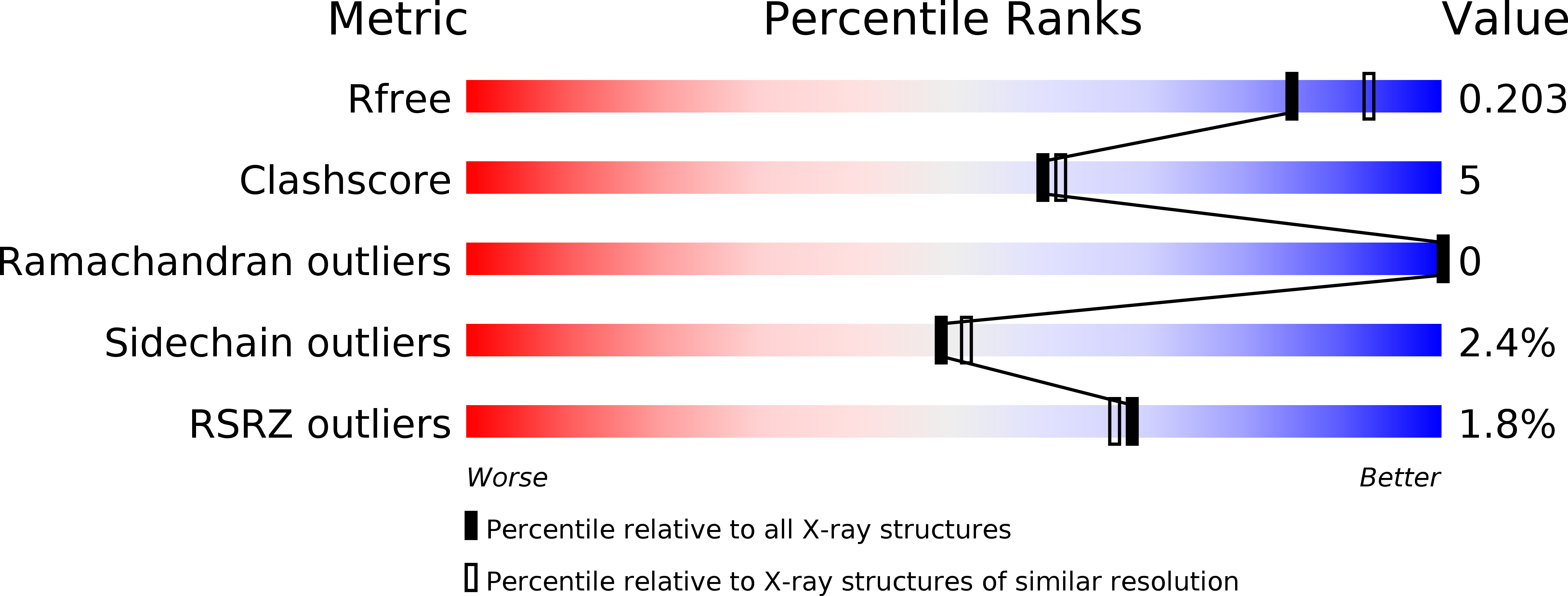
2.00 Å
R-Value Free:
0.19
R-Value Work:
0.15
Space Group:
P 1
Macromolecular Entities
Polymer Type:polypeptide(L)
Molecule:PENICILLIN G ACYLASE ALPHA SUBUNIT
Gene (Uniprot):pac
Chain IDs:A
Chain Length:209
Number of Molecules:1
Biological Source:ESCHERICHIA COLI
Polymer Type:polypeptide(L)
Molecule:PENICILLIN G ACYLASE BETA SUBUNIT
Gene (Uniprot):pac
Mutations:YES
Chain IDs:B
Chain Length:557
Number of Molecules:1
Biological Source:ESCHERICHIA COLI
Primary Citation
Mutations of Penicillin Acylase Residue B71 Extend Substrate Specificity by Decreasing Steric Constraints for Substrate Binding
Biochem.J. 371 143 ? (2003)
PMID: 12511194 DOI: 10.1042/BJ20021383

Abstact

Two mutant forms of penicillin acylase from Escherichia coli strains, selected using directed evolution for the ability to use glutaryl-L-leucine for growth [Forney, Wong and Ferber (1989) Appl. Environ. Microbiol. 55, 2550-2555], are changed within one codon, replacing the B-chain residue Phe(B71) with either Cys or Leu. Increases of up to a factor of ten in k (cat)/ K (m) values for substrates possessing a phenylacetyl leaving group are consistent with a decrease in K (s). Values of k (cat)/ K (m) for glutaryl-L-leucine are increased at least 100-fold. A decrease in k (cat)/ K (m) for the Cys(B71) mutant with increased pH is consistent with binding of the uncharged glutaryl group. The mutant proteins are more resistant to urea denaturation monitored by protein fluorescence, to inactivation in the presence of substrate either in the presence of urea or at high pH, and to heat inactivation. The crystal structure of the Leu(B71) mutant protein, solved to 2 A resolution, shows a flip of the side chain of Phe(B256) into the periphery of the catalytic centre, associated with loss of the pi-stacking interactions between Phe(B256) and Phe(B71). Molecular modelling demonstrates that glutaryl-L-leucine may bind with the uncharged glutaryl group in the S(1) subsite of either the wild-type or the Leu(B71) mutant but with greater potential freedom of rotation of the substrate leucine moiety in the complex with the mutant protein. This implies a smaller decrease in the conformational entropy of the substrate on binding to the mutant proteins and consequently greater catalytic activity.

Legend

Protein

Chemical

Disease

Primary Citation of related structures