Deposition Date
2002-07-21
Release Date
2003-07-07
Last Version Date
2023-12-13
Entry Detail
PDB ID:
1H1T
Keywords:
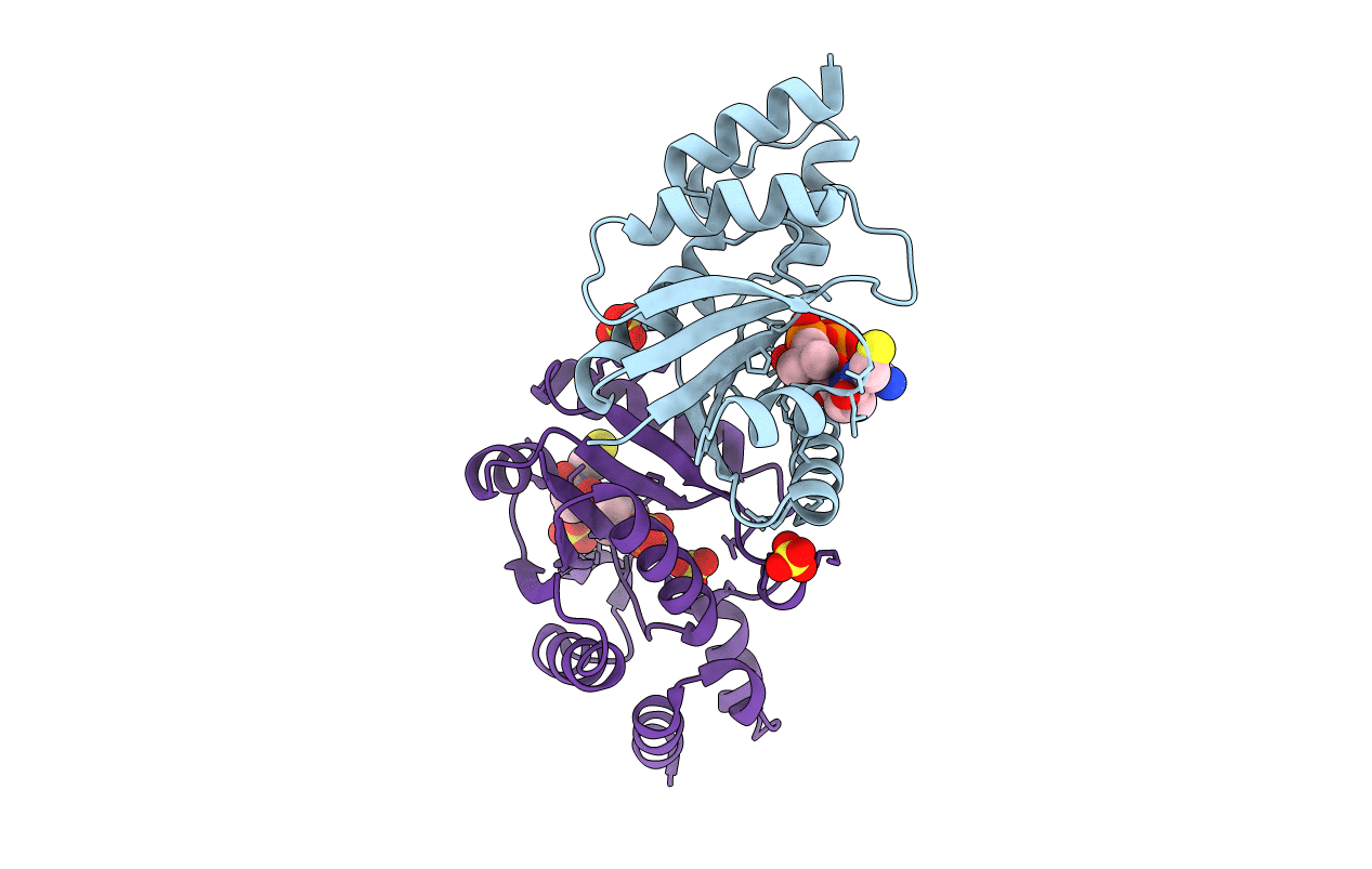
Title:
PHOSPHOPANTETHEINE ADENYLYLTRANSFERASE IN COMPLEX WITH Coenzyme A FROM ESCHERICHIA COLI
Biological Source:
Source Organism(s):
ESCHERICHIA COLI (Taxon ID: 562)
Method Details:
Experimental Method:
Resolution:
1.78 Å
R-Value Free:
0.24
R-Value Work:
0.21
R-Value Observed:
0.21
Space Group:
I 2 3