1H1N image
Deposition Date 2002-07-19
Release Date 2002-08-12
Last Version Date 2024-11-06
Entry Detail
PDB ID:
1H1N
Keywords:
Title:
Atomic resolution structure of the major endoglucanase from Thermoascus aurantiacus
Biological Source:
Source Organism:
Method Details:
Experimental Method:
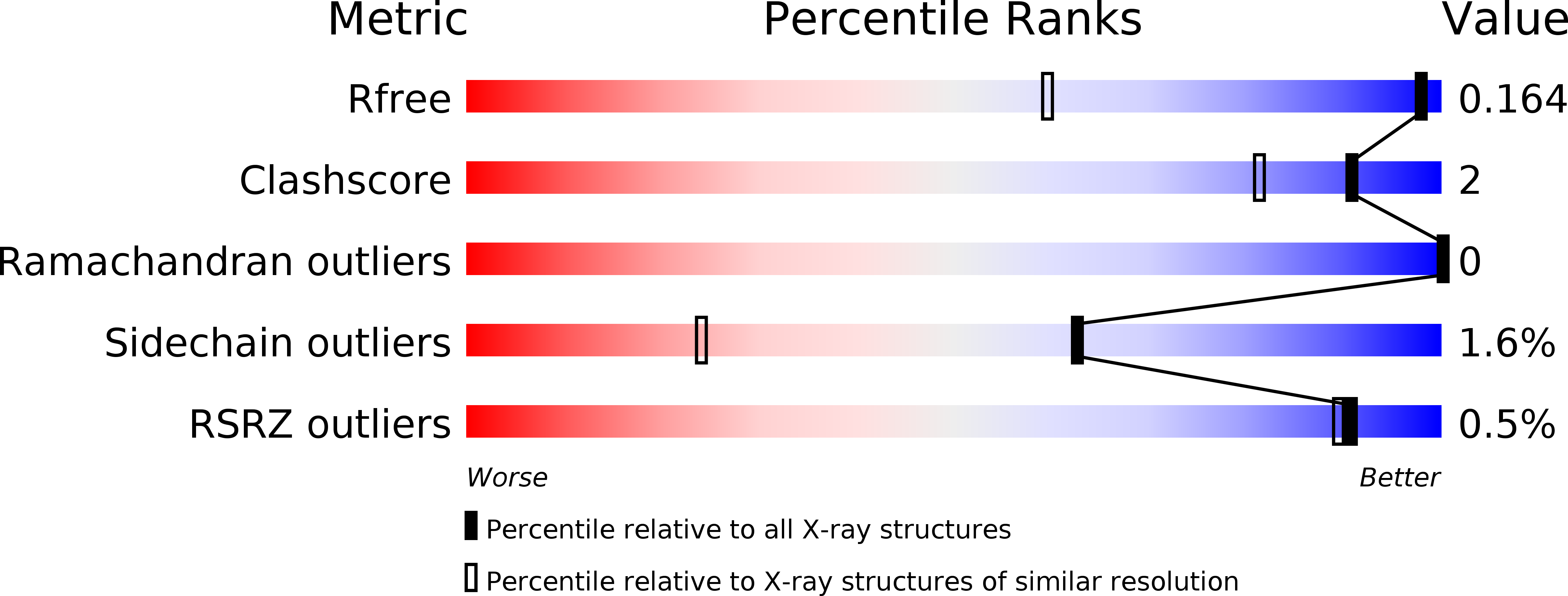
Resolution:
1.12 Å
R-Value Free:
0.17
R-Value Observed:
0.14
Space Group:
P 21 21 21
Macromolecular Entities
Structures with similar UniProt ID
Protein Blast
Polymer Type:polypeptide(L)
Molecule:ENDO TYPE CELLULASE ENGI
Gene (Uniprot):eg1
Chain IDs:A, B
Chain Length:305
Number of Molecules:2
Biological Source:THERMOASCUS AURANTIACUS
Primary Citation
Atomic Resolution Structure of the Major Endoglucanase from Thermoascus Aurantiacus
Biochem.Biophys.Res.Commun. 296 161 ? (2002)
PMID: 12147244 DOI: 10.1016/S0006-291X(02)00775-1

Abstact

The crystal structure of the major endoglucanase from the thermophilic fungus Thermoascus aurantiacus was determined by single isomorphous replacement at 1.12A resolution. The full sequence supports the classification of the protein in a subgroup of glycoside hydrolase family 5 for which no structural data are available yet. The active site shows eight critical residues, strictly conserved within family 5. In addition, aromatic residues that line the substrate-binding cleft and that are possibly involved in substrate-binding are identified. A number of residues seem to be conserved among members of the subtype, including a disulphide bridge between Cys212 and Cys249.

Legend

Protein

Chemical

Disease

Primary Citation of related structures
Feedback Form
Name
Email
Institute
Feedback