Deposition Date
2002-05-24
Release Date
2003-11-20
Last Version Date
2024-10-23
Entry Detail
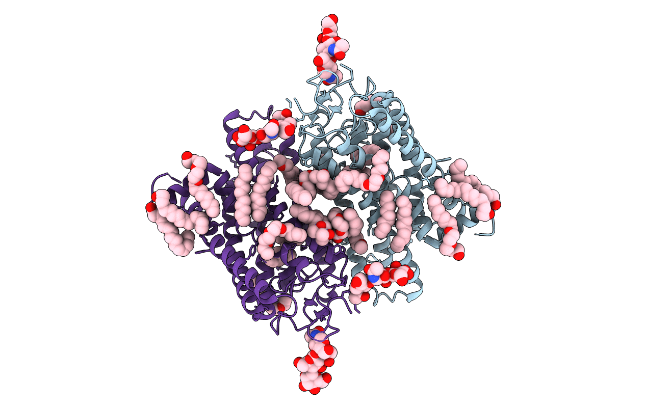
PDB ID:
1GZM
Keywords:
Title:
Structure of Bovine Rhodopsin in a Trigonal Crystal Form
Biological Source:
Source Organism(s):
BOS TAURUS (Taxon ID: 9913)
Method Details:
Experimental Method:
Resolution:
2.65 Å
R-Value Free:
0.23
R-Value Work:
0.20
R-Value Observed:
0.20
Space Group:
P 31