1GZ6 image
Deposition Date 2002-05-16
Release Date 2003-01-24
Last Version Date 2024-11-06
Entry Detail
PDB ID:
1GZ6
Keywords:
Title:
(3R)-HYDROXYACYL-COA DEHYDROGENASE FRAGMENT OF RAT PEROXISOMAL MULTIFUNCTIONAL ENZYME TYPE 2
Biological Source:
Source Organism:
Host Organism:
Method Details:
Experimental Method:
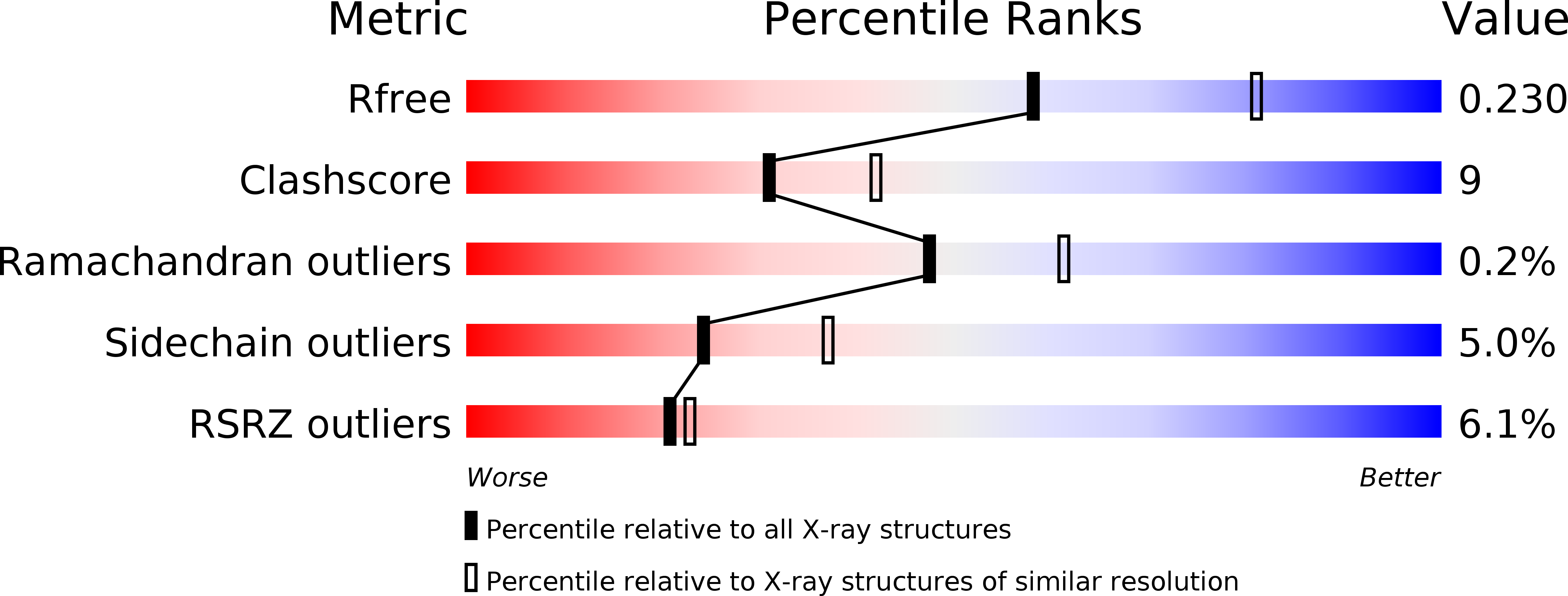
Resolution:
2.38 Å
R-Value Free:
0.23
R-Value Work:
0.19
R-Value Observed:
0.19
Space Group:
P 1 21 1
Macromolecular Entities
Polymer Type:polypeptide(L)
Molecule:ESTRADIOL 17 BETA-DEHYDROGENASE 4
Gene (Uniprot):Hsd17b4
Chain IDs:A, B, C, D
Chain Length:319
Number of Molecules:4
Biological Source:RATTUS NORVEGICUS
Modified Residue
Compound ID Chain ID Parent Comp ID Details 2D Image
MSE A MET SELENOMETHIONINE
Primary Citation
Binary Structure of the Two-Domain (3R)-Hydroxyacyl-Coa Dehydrogenase from Rat Peroxisomal Multifunctional Enzyme Type 2 at 2.38 A Resolution
Structure 11 87 ? (2003)
PMID: 12517343 DOI: 10.1016/S0969-2126(02)00931-0

Abstact

The crystal structure of (3R)-hydroxyacyl-CoA dehydrogenase of rat peroxisomal multifunctional enzyme type 2 (MFE-2) was solved at 2.38 A resolution. The catalytic entity reveals an alpha/beta short chain alcohol dehydrogenase/reductase (SDR) fold and the conformation of the bound nicotinamide adenine dinucleotide (NAD(+)) found in other SDR enzymes. Of great interest is the separate COOH-terminal domain, which is not seen in other SDR structures. This domain completes the active site cavity of the neighboring monomer and extends dimeric interactions. Peroxisomal diseases that arise because of point mutations in the dehydrogenase-coding region of the MFE-2 gene can be mapped to changes in amino acids involved in NAD(+) binding and protein dimerization.

Legend

Protein

Chemical

Disease

Primary Citation of related structures