1GXS image
Deposition Date 2002-04-11
Release Date 2002-10-01
Last Version Date 2024-10-16
Entry Detail
PDB ID:
1GXS
Keywords:
Title:
Crystal Structure of Hydroxynitrile Lyase from Sorghum bicolor in Complex with Inhibitor Benzoic Acid: a novel cyanogenic enzyme
Biological Source:
Source Organism:
SORGHUM BICOLOR (Taxon ID: 4558)
Method Details:
Experimental Method:
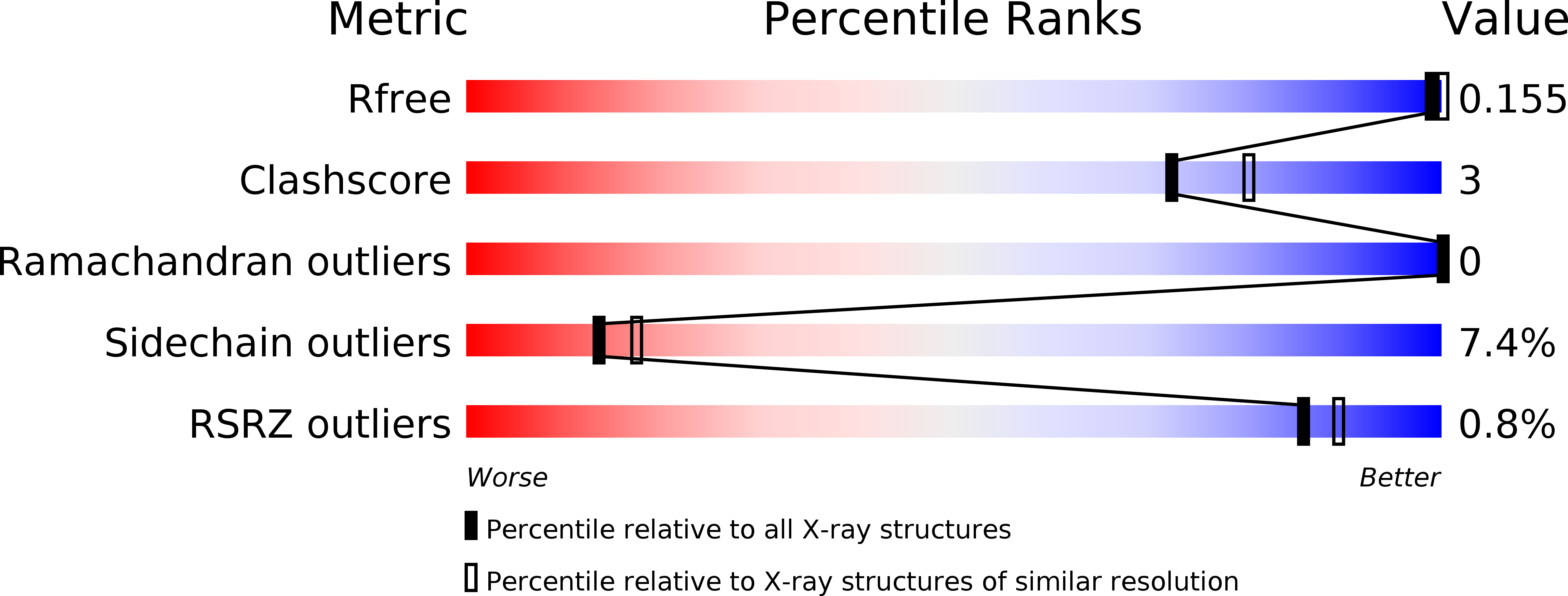
Resolution:
2.30 Å
R-Value Free:
0.22
R-Value Work:
0.16
R-Value Observed:
0.16
Space Group:
C 1 2 1
Macromolecular Entities
Structures with similar UniProt ID
Protein Blast
Polymer Type:polypeptide(L)
Molecule:P-(S)-HYDROXYMANDELONITRILE LYASE CHAIN A
Chain IDs:A, C
Chain Length:270
Number of Molecules:2
Biological Source:SORGHUM BICOLOR
Structures with similar UniProt ID
Protein Blast
Polymer Type:polypeptide(L)
Molecule:P-(S)-HYDROXYMANDELONITRILE LYASE CHAIN B
Chain IDs:B, D
Chain Length:158
Number of Molecules:2
Biological Source:SORGHUM BICOLOR
Modified Residue
Compound ID Chain ID Parent Comp ID Details 2D Image
ASN A ASN GLYCOSYLATION SITE
Primary Citation
Crystal Structure of Hydroxynitrile Lyase from Sorghum Bicolor in Complex with the Inhibitor Benzoic Acid: A Novel Cyanogenic Enzyme
Biochemistry 41 12043 ? (2002)
PMID: 12356304 DOI: 10.1021/BI020300O

Abstact

The crystal structure of the hydroxynitrile lyase from Sorghum bicolor (SbHNL) in complex with the inhibitor benzoic acid has been determined at 2.3 A resolution and refined to a crystallographic R-factor of 16.5%. The SbHNL sequence places the enzyme in the alpha/beta hydrolase family where the active site nucleophile is predicted to be organized in a characteristic pentapeptide motif which is part of the active site strand-turn-helix motif. In SbHNL, however, a unique two-amino acid deletion is next to the putative active site Ser158, removing thereby the putative oxyanion hole-forming Tyr residue. The presented X-ray structure shows that the overall folding pattern of SbHNL is similar to that of the closely related wheat serine carboxypeptidase (CPD-WII); however, the deletion in SbHNL is forcing the putative active site residues away from the expected hydrolase binding site toward a small hydrophobic cleft, which also contains the inhibitor benzoic acid, defining thereby a completely different SbHNL active site architecture where the traditional view of a classic triad is not given any more. Rather, we propose a mechanism involving general base catalysis by the carboxy-terminal Trp270 carboxyl group and proton transfer toward the leaving nitrile group by an active site water molecule. The unexpected interactions of the inhibitor with the new SbHNL active site also reveal the structural basis for the enzyme's limited substrate specificity. The implications of this structure on the evolution of catalysis in the hydroxynitrile lyase superfamily are discussed.

Legend

Protein

Chemical

Disease

Primary Citation of related structures