1GW3 image
Deposition Date 1997-06-04
Release Date 1997-07-23
Last Version Date 2024-05-22
Entry Detail
PDB ID:
1GW3
Title:
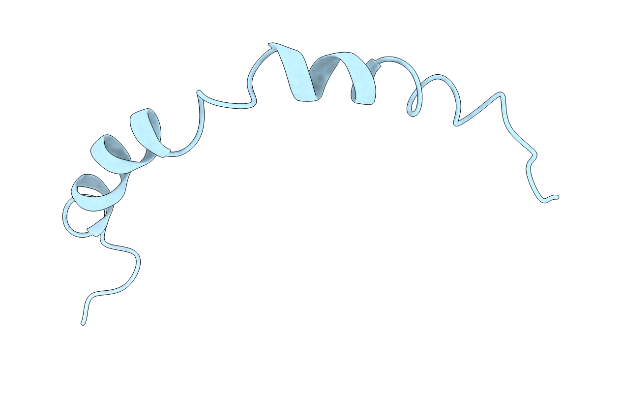
THE HELIX-HINGE-HELIX STRUCTURAL MOTIF IN HUMAN APOLIPOPROTEIN A-I DETERMINED BY NMR SPECTROSCOPY, 1 STRUCTURE
Biological Source:
Source Organism:
Homo sapiens (Taxon ID: 9606)
Method Details:
Experimental Method:
Conformers Calculated:
50
Conformers Submitted:
1
Selection Criteria:
LOWER OPTIMIZATION ERROR AND NO DISTANCE VIOLATION GREATER THAN 0.5 A)
Macromolecular Entities
Polymer Type:polypeptide(L)
Molecule:APOA-I
Gene (Uniprot):APOA1
Chain IDs:A
Chain Length:46
Number of Molecules:1
Biological Source:Homo sapiens
Ligand Molecules
Primary Citation
The helix-hinge-helix structural motif in human apolipoprotein A-I determined by NMR spectroscopy.
Biochemistry 36 13657 13666 (1997)
PMID: 9354635 DOI: 10.1021/bi971151q

Abstact

The conformation of a synthetic peptide of 46 residues from apoA-I was investigated by fluorescence, CD, and 2D NMR spectroscopies in lipid-mimetic environments. ApoA-I(142-187) is mainly unstructured in water but helical in SDS or dodecylphosphocholine (DPC), although the peptide only associates with DPC at approximately the critical micellar concentration. Solution structures of apoA-I(142-187) were determined by distance geometry calculations based on 450 (in DPC-d38) or 397 (in SDS-d25) NOE-derived distance restraints, respectively. Backbone RMSDs for superimposing the two helical regions 146-162 and 168-182 are 0.98 +/- 0.22 (2.38 +/- 0.20) and 1.99 +/- 0.42 (2.02 +/- 0.21) A in DPC (SDS), respectively. No interhelical NOE was found, suggesting that helix-helix interactions between the two helical domains in apoA-I(142-187) are unlikely. Similar average, curved helix-hinge-helix structures were found in both SDS and DPC micelles with the hydrophobic residues occupying the concave face, indicating that hydrophobic interactions dominate. Intermolecular NOESY experiments, performed in the presence of 50% protonated SDS, confirm that the two amphipathic helices and Y166 in the hinge all interact with the micelle. The involvement of Y166 in lipid binding is supported by fluorescence spectroscopy as well. On the basis of all the data above, we propose a model for the peptide-lipid complexes wherein the curved amphipathic helix-hinge-helix structural motif straddles the micelle. The peptide-aided signal assignment achieved for apoA-I(122-187) (66mer) and apoA-I suggests that such a structural motif is retained in the longer peptide and most likely in the intact protein.

Legend

Protein

Chemical

Disease

Primary Citation of related structures