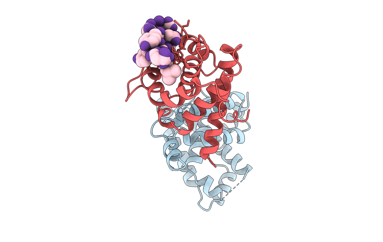
1GUX image
Deposition Date 1997-11-15
Release Date 1998-12-02
Last Version Date 2024-02-07
Entry Detail
PDB ID:
1GUX
Title:
RB POCKET BOUND TO E7 LXCXE MOTIF
Biological Source:
Source Organism:
Method Details:
Experimental Method:
Resolution:
1.85 Å
R-Value Free:
0.28
R-Value Work:
0.21
R-Value Observed:
0.21
Space Group:
C 1 2 1
Macromolecular Entities
Structures with similar UniProt ID
Protein Blast
Polymer Type:polypeptide(L)
Molecule:RETINOBLASTOMA PROTEIN
Gene (Uniprot):RB1
Chain IDs:A
Chain Length:218
Number of Molecules:1
Biological Source:Homo sapiens
Structures with similar UniProt ID
Protein Blast
Polymer Type:polypeptide(L)
Molecule:RETINOBLASTOMA PROTEIN
Gene (Uniprot):RB1
Chain IDs:B
Chain Length:152
Number of Molecules:1
Biological Source:Homo sapiens
Polymer Type:polypeptide(L)
Molecule:ONCOPROTEIN
Chain IDs:C (auth: E)
Chain Length:9
Number of Molecules:1
Biological Source:Human papillomavirus
Primary Citation
Structure of the retinoblastoma tumour-suppressor pocket domain bound to a peptide from HPV E7.
Nature 391 859 865 (1998)
PMID: 9495340 DOI: 10.1038/36038

Abstact

The pocket domain of the retinoblastoma (Rb) tumour suppressor is central to Rb function, and is frequently inactivated by the binding of the human papilloma virus E7 oncoprotein in cervical cancer. The crystal structure of the Rb pocket bound to a nine-residue E7 peptide containing the LxCxE motif, shared by other Rb-binding viral and cellular proteins, shows that the LxCxE peptide binds a highly conserved groove on the B-box portion of the pocket; the A-box portion appears to be required for the stable folding of the B box. Also highly conserved is the extensive A-B interface, suggesting that it may be an additional protein-binding site. The A and B boxes each contain the cyclin-fold structural motif, with the LxCxE-binding site on the B-box cyclin fold being similar to a Cdk2-binding site of cyclin A and to a TBP-binding site of TFIIB.

Legend

Protein

Chemical

Disease

Primary Citation of related structures