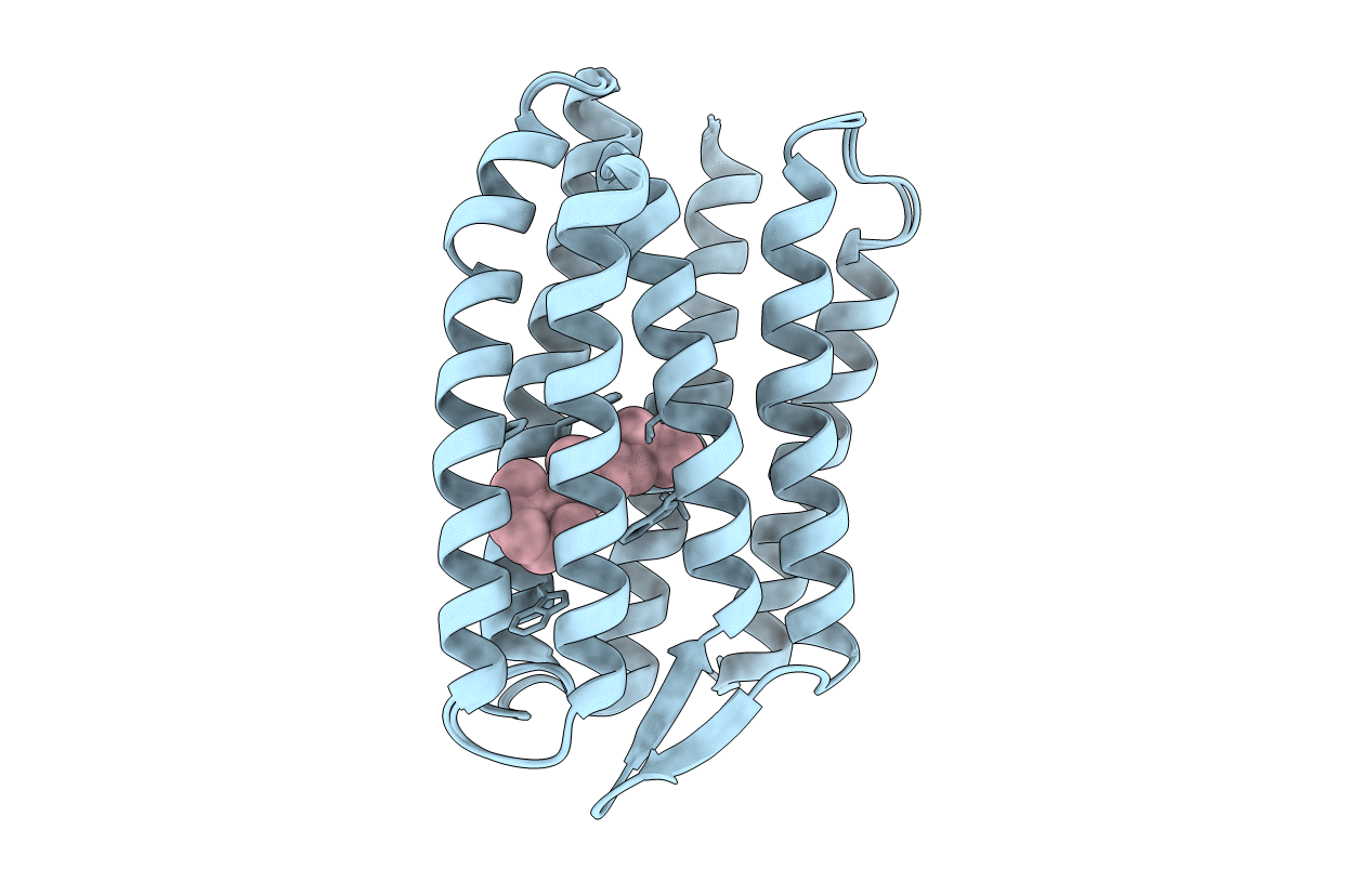
1GU8 image
Entry Detail
PDB ID:
1GU8
Title:
SENSORY RHODOPSIN II
Biological Source:
Source Organism:
Host Organism:
PDB Version:
Deposition Date:
2002-01-24
Release Date:
2002-04-12
Method Details:
Experimental Method:
Resolution:
2.27 Å
R-Value Free:
0.25
R-Value Work:
0.23
R-Value Observed:
0.23
Space Group:
C 2 2 21
Macromolecular Entities
Polymer Type:polypeptide(L)
Description:SENSORY RHODOPSIN II
Chain IDs:A
Chain Length:239
Number of Molecules:1
Biological Source:NATRONOMONAS PHARAONIS
Primary Citation
Early Structural Rearrangements in the Photocycle of an Integral Membrane Sensory Receptor
Structure 10 473 ? (2002)
PMID: 11937052 DOI: 10.1016/S0969-2126(02)00736-0

Abstact

Sensory rhodopsins are the primary receptors of vision in animals and phototaxis in microorganisms. Light triggers the rapid isomerization of a buried retinal chromophore, which the protein both accommodates and amplifies into the larger structural rearrangements required for signaling. We trapped an early intermediate of the photocycle of sensory rhodopsin II from Natronobacterium pharaonis (pSRII) in 3D crystals and determined its X-ray structure to 2.3 A resolution. The observed structural rearrangements were localized near the retinal chromophore, with a key water molecule becoming disordered and the retinal's beta-ionone ring undergoing a prominent movement. Comparison with the early structural rearrangements of bacteriorhodopsin illustrates how modifications in the retinal binding pocket of pSRII allow subtle differences in the early relaxation of photoisomerized retinal.

Legend

Protein

Chemical

Disease

Primary Citation of related structures