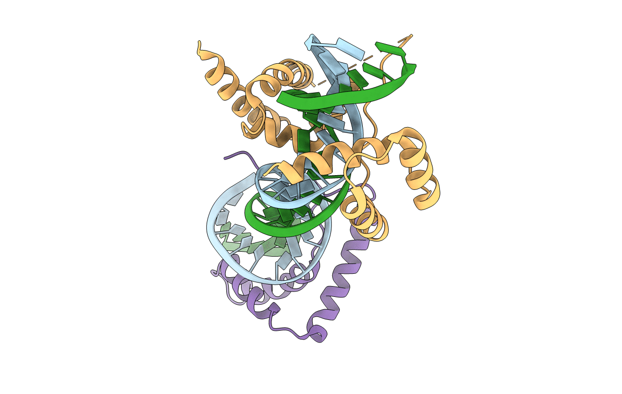
1GT0 image
Entry Detail
PDB ID:
1GT0
Keywords:
Title:
Crystal structure of a POU/HMG/DNA ternary complex
Biological Source:
Source Organism:
Host Organism:
PDB Version:
Deposition Date:
2002-01-09
Release Date:
2003-01-30
Method Details:
Experimental Method:
Resolution:
2.60 Å
R-Value Free:
0.28
R-Value Work:
0.23
R-Value Observed:
0.23
Space Group:
P 31 2 1
Macromolecular Entities
Polymer Type:polydeoxyribonucleotide
Description:5'-D(*TP*TP*CP*TP*TP*TP*GP*TP*TP*TP* GP*GP*AP* TP*GP*CP*TP*AP*AP*TP*GP*GP*GP*A)-3'
Chain IDs:A
Chain Length:24
Number of Molecules:1
Biological Source:MUS MUSCULUS
Polymer Type:polydeoxyribonucleotide
Description:5'-D(*AP*TP*CP*CP*CP*AP*TP*TP*AP*GP* CP*AP*TP*CP*CP*AP*AP*AP*CP*AP*AP*AP*GP*A)-3'
Chain IDs:B
Chain Length:24
Number of Molecules:1
Biological Source:MUS MUSCULUS
Polymer Type:polypeptide(L)
Description:OCTAMER-BINDING TRANSCRIPTION FACTOR 1
Mutations:YES
Chain IDs:C
Chain Length:159
Number of Molecules:1
Biological Source:HOMO SAPIENS
Polymer Type:polypeptide(L)
Description:TRANSCRIPTION FACTOR SOX-2
Chain IDs:D
Chain Length:80
Number of Molecules:1
Biological Source:MUS MUSCULUS
Primary Citation
Crystal Structure of a POU/Hmg/DNA Ternary Complex Suggests Differential Assembly of Oct4 and Sox2 on Two Enhancers
Genes Dev. 17 2048 ? (2003)
PMID: 12923055 DOI: 10.1101/GAD.269303

Abstact

Members of the POU and SOX transcription factor families exemplify the partnerships established between various transcriptional regulators during early embryonic development. Although functional cooperativity between key regulator proteins is pivotal for milestone decisions in mammalian development, little is known about the underlying molecular mechanisms. In this study, we focus on two transcription factors, Oct4 and Sox2, as their combination on DNA is considered to direct the establishment of the first three lineages in the mammalian embryo. Using experimental high-resolution structure determination, followed by model building and experimental validation, we found that Oct4 and Sox2 were able to dimerize onto DNA in distinct conformational arrangements. We demonstrate that the DNA enhancer region of their target genes is responsible for the correct spatial alignment of glue-like interaction domains on their surface. Interestingly, these surfaces frequently have redundant functions and are instrumental in recruiting various interacting protein partners.

Legend

Protein

Chemical

Disease

Primary Citation of related structures