Deposition Date
2001-12-14
Release Date
2002-05-16
Last Version Date
2024-11-06
Entry Detail
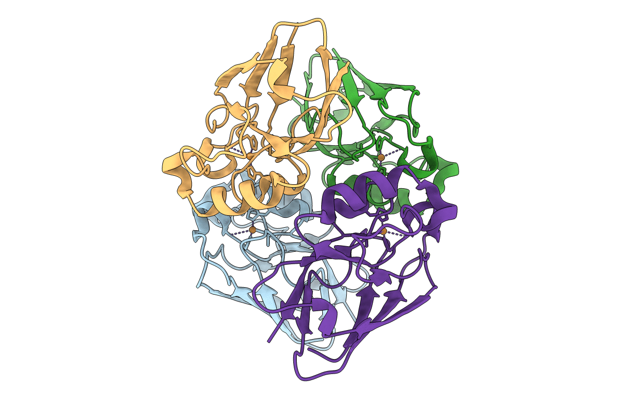
PDB ID:
1GR7
Keywords:
Title:
Crystal structure of the double mutant Cys3Ser/Ser100Pro from Pseudomonas Aeruginosa at 1.8 A resolution
Biological Source:
Source Organism(s):
PSEUDOMONAS AERUGINOSA (Taxon ID: 287)
Expression System(s):
Method Details:
Experimental Method:
Resolution:
1.80 Å
R-Value Free:
0.20
R-Value Work:
0.17
R-Value Observed:
0.17
Space Group:
P 21 21 21