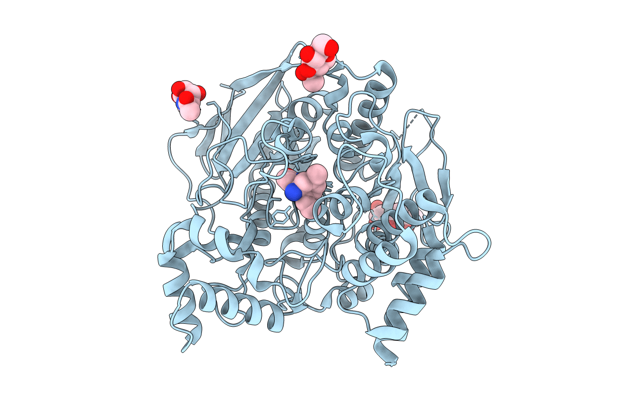
1GPK image
Deposition Date 2001-11-05
Release Date 2002-08-29
Last Version Date 2024-10-16
Entry Detail
PDB ID:
1GPK
Keywords:
Title:
Structure of Acetylcholinesterase Complex with (+)-Huperzine A at 2.1A Resolution
Biological Source:
Source Organism:
Method Details:
Experimental Method:
Resolution:
2.10 Å
R-Value Free:
0.21
R-Value Work:
0.18
R-Value Observed:
0.18
Space Group:
P 31 2 1
Macromolecular Entities
Polymer Type:polypeptide(L)
Molecule:ACETYLCHOLINESTERASE
Gene (Uniprot):ache
Chain IDs:A
Chain Length:537
Number of Molecules:1
Biological Source:TORPEDO CALIFORNICA
Modified Residue
Compound ID Chain ID Parent Comp ID Details 2D Image
ASN A ASN GLYCOSYLATION SITE
Primary Citation
X-Ray Structures of Torpedo Californica Acetylcholinesterase Complexed with (+)-Huperzine a and (-)-Huperzine B: Structural Evidence for an Active Site Rearrangement
Biochemistry 41 10810 ? (2002)
PMID: 12196020 DOI: 10.1021/BI020151+

Abstact

Kinetic and structural data are presented on the interaction with Torpedo californica acetylcholinesterase (TcAChE) of (+)-huperzine A, a synthetic enantiomer of the anti-Alzheimer drug, (-)-huperzine A, and of its natural homologue (-)-huperzine B. (+)-Huperzine A and (-)-huperzine B bind to the enzyme with dissociation constants of 4.30 and 0.33 microM, respectively, compared to 0.18 microM for (-)-huperzine A. The X-ray structures of the complexes of (+)-huperzine A and (-)-huperzine B with TcAChE were determined to 2.1 and 2.35 A resolution, respectively, and compared to the previously determined structure of the (-)-huperzine A complex. All three interact with the "anionic" subsite of the active site, primarily through pi-pi stacking and through van der Waals or C-H.pi interactions with Trp84 and Phe330. Since their alpha-pyridone moieties are responsible for their key interactions with the active site via hydrogen bonding, and possibly via C-H.pi interactions, all three maintain similar positions and orientations with respect to it. The carbonyl oxygens of all three appear to repel the carbonyl oxygen of Gly117, thus causing the peptide bond between Gly117 and Gly118 to undergo a peptide flip. As a consequence, the position of the main chain nitrogen of Gly118 in the "oxyanion" hole in the native enzyme becomes occupied by the carbonyl of Gly117. Furthermore, the flipped conformation is stabilized by hydrogen bonding of Gly117O to Gly119N and Ala201N, the other two functional elements of the three-pronged "oxyanion hole" characteristic of cholinesterases. All three inhibitors thus would be expected to abolish hydrolysis of all ester substrates, whether charged or neutral.

Legend

Protein

Chemical

Disease