1GO3 image
Deposition Date 2001-10-17
Release Date 2001-12-07
Last Version Date 2024-05-08
Entry Detail
PDB ID:
1GO3
Keywords:
Title:
Structure of an archeal homolog of the eukaryotic RNA polymerase II RPB4/RPB7 complex
Biological Source:
Source Organism:
Method Details:
Experimental Method:
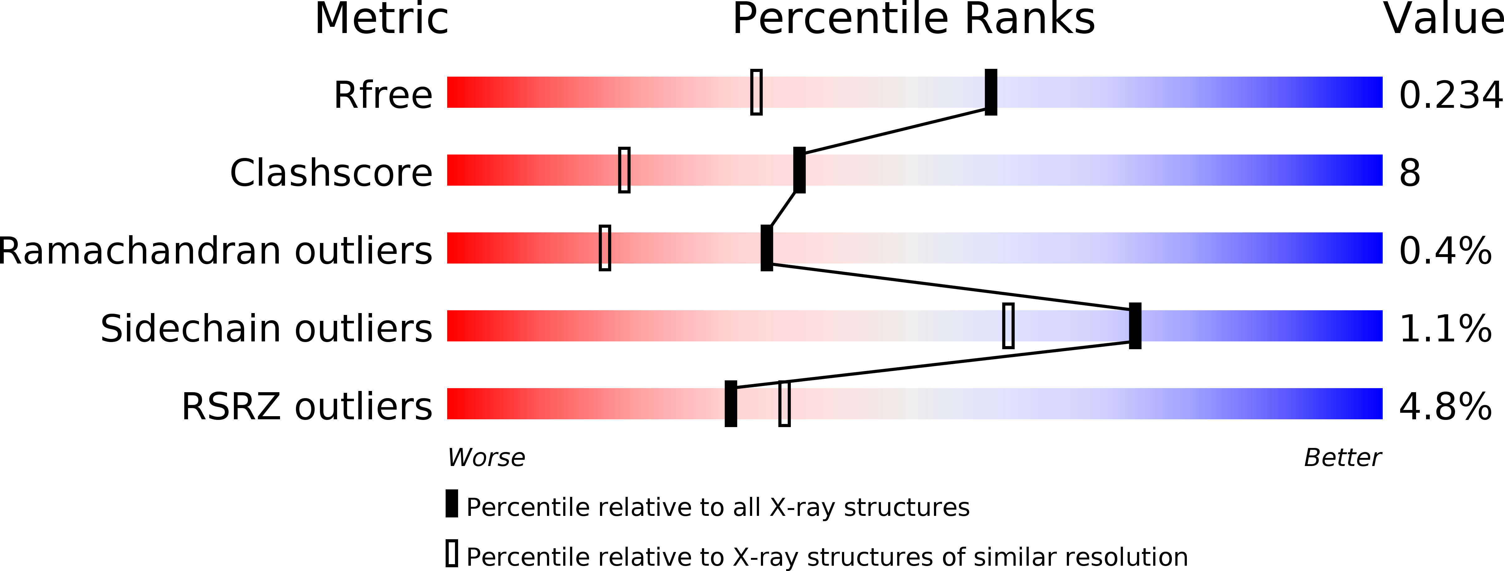
Resolution:
1.75 Å
R-Value Free:
0.23
R-Value Work:
0.21
R-Value Observed:
0.21
Space Group:
P 43
Macromolecular Entities
Polymer Type:polypeptide(L)
Molecule:DNA-DIRECTED RNA POLYMERASE SUBUNIT E
Gene (Uniprot):rpo7
Chain IDs:A (auth: E), C (auth: M)
Chain Length:187
Number of Molecules:2
Biological Source:METHANOCOCCUS JANNASCHII
Polymer Type:polypeptide(L)
Molecule:DNA-DIRECTED RNA POLYMERASE SUBUNIT F
Gene (Uniprot):rpo4
Chain IDs:B (auth: F), D (auth: N)
Chain Length:107
Number of Molecules:2
Biological Source:METHANOCOCCUS JANNASCHII
Primary Citation
Structure of an Archaeal Homolog of the Eukaryotic RNA Polymerase II Rpb4/Rpb7 Complex
Mol.Cell 8 1137 1143 (2001)
PMID: 11741548 DOI: 10.1016/S1097-2765(01)00379-3

Abstact

The eukaryotic subunits RPB4 and RPB7 form a heterodimer that reversibly associates with the RNA polymerase II core and constitute the only two components of the enzyme for which no structural information is available. We have determined the crystal structure of the complex between the Methanococcus jannaschii subunits E and F, the archaeal homologs of RPB7 and RPB4. Subunit E has an elongated two-domain structure and contains two potential RNA binding motifs, while the smaller F subunit wraps around one side of subunit E, at the interface between the two domains. We propose a model for the interaction between RPB4/RPB7 and the core RNA polymerase in which the RNA binding face of RPB7 is positioned to interact with the nascent RNA transcript.

Legend

Protein

Chemical

Disease

Primary Citation of related structures