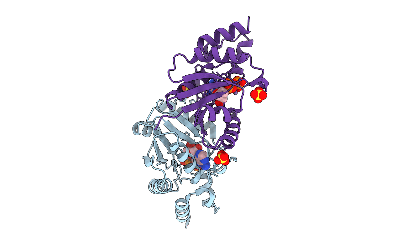
1GN8 image
Deposition Date 2001-10-03
Release Date 2002-01-17
Last Version Date 2023-12-13
Entry Detail
PDB ID:
1GN8
Keywords:
Title:
PHOSPHOPANTETHEINE ADENYLYLTRANSFERASE IN COMPLEX WITH Mn2+ATP FROM ESCHERICHIA COLI
Biological Source:
Source Organism:
ESCHERICHIA COLI (Taxon ID: 562)
Method Details:
Experimental Method:
Resolution:
1.83 Å
R-Value Free:
0.24
R-Value Work:
0.22
R-Value Observed:
0.22
Space Group:
I 2 3
Macromolecular Entities
Structures with similar UniProt ID
Protein Blast
Polymer Type:polypeptide(L)
Molecule:PHOSPHOPANTETHEINE ADENYLYLTRANSFERASE
Chain IDs:A, B
Chain Length:159
Number of Molecules:2
Biological Source:ESCHERICHIA COLI
Primary Citation
The Crystal Structures of Phosphopantetheine Adenylyltransferase with Bound Substrates Reveal the Enzyme'S Catalytic Mechanism
J.Mol.Biol. 315 487 ? (2001)
PMID: 11812124 DOI: 10.1006/JMBI.2001.5272

Abstact

Phosphopantetheine adenylyltransferase (PPAT) is an essential enzyme in the coenzyme A pathway that catalyzes the reversible transfer of an adenylyl group from ATP to 4'-phosphopantetheine (Ppant) in the presence of magnesium. To investigate the reaction mechanism, the high-resolution crystal structures of the Escherichia coli PPAT have been determined in the presence of either ATP or Ppant. Structural details of the catalytic center revealed specific roles for individual amino acid residues involved in substrate binding and catalysis. The side-chain of His18 stabilizes the expected pentacovalent intermediate, whereas the side-chains of Thr10 and Lys42 orient the nucleophile for an in-line displacement mechanism. The binding site for the manganese ion that interacts with the phosphate groups of the nucleotide has also been identified. Within the PPAT hexamer, one trimer is in its substrate-free state, whereas the other is in a substrate-bound state.

Legend

Protein

Chemical

Disease

Primary Citation of related structures