Deposition Date
2001-09-12
Release Date
2001-11-28
Last Version Date
2023-12-13
Entry Detail
PDB ID:
1GM9
Keywords:
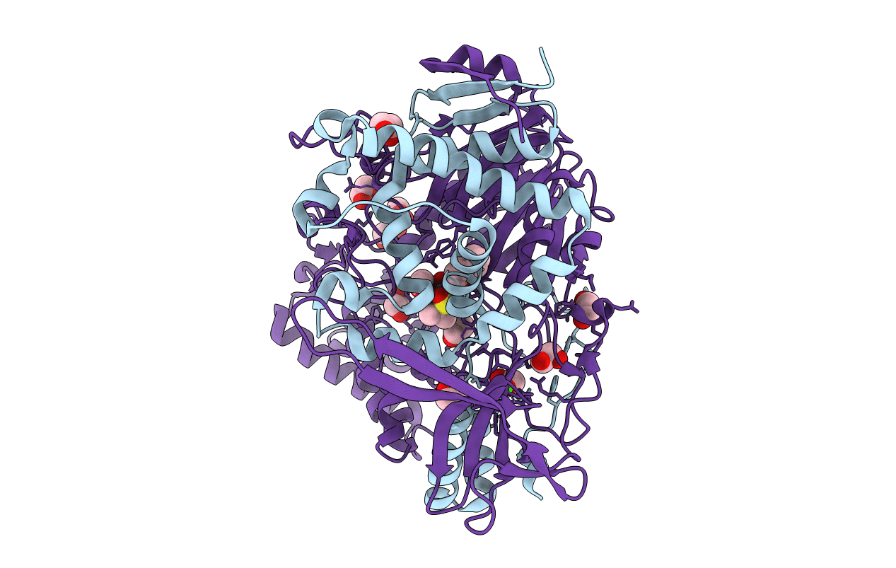
Title:
Crystal structures of penicillin acylase enzyme-substrate complexes: Structural insights into the catalytic mechanism
Biological Source:
Source Organism(s):
ESCHERICHIA COLI (Taxon ID: 562)
Expression System(s):
Method Details:
Experimental Method:
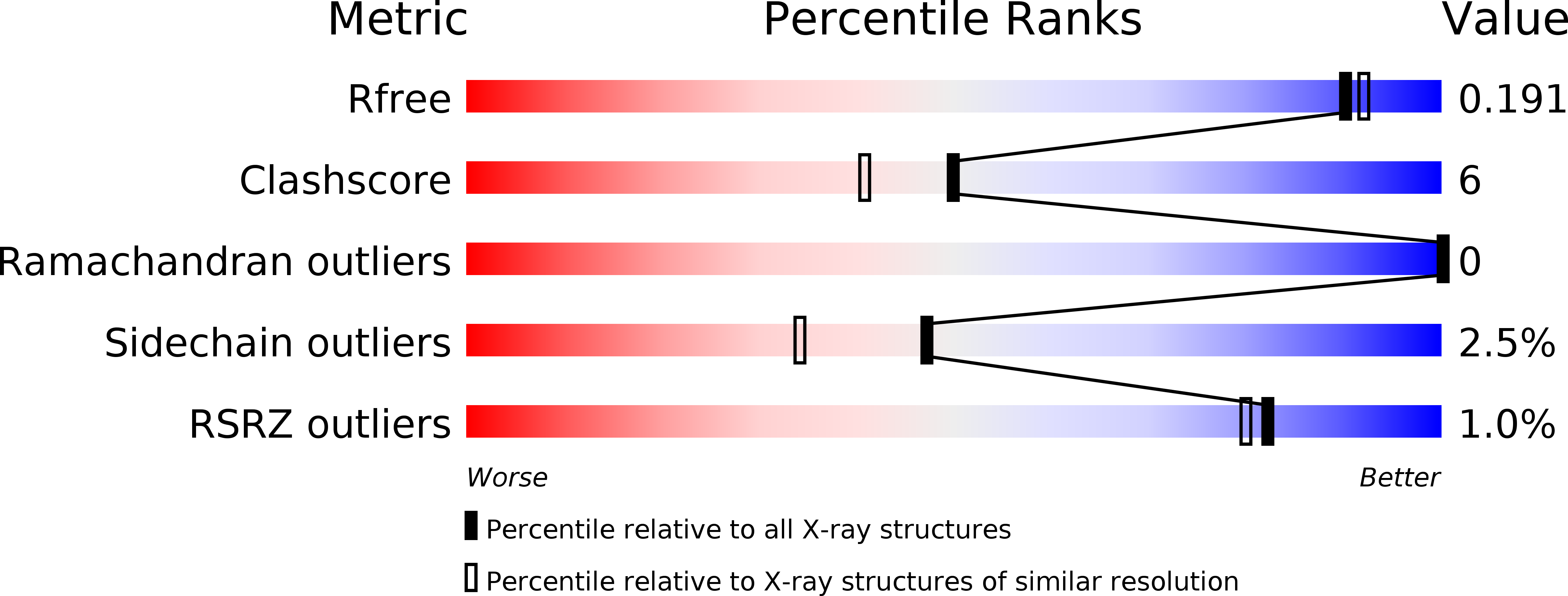
Resolution:
1.80 Å
R-Value Free:
0.19
R-Value Work:
0.15
Space Group:
P 1 21 1