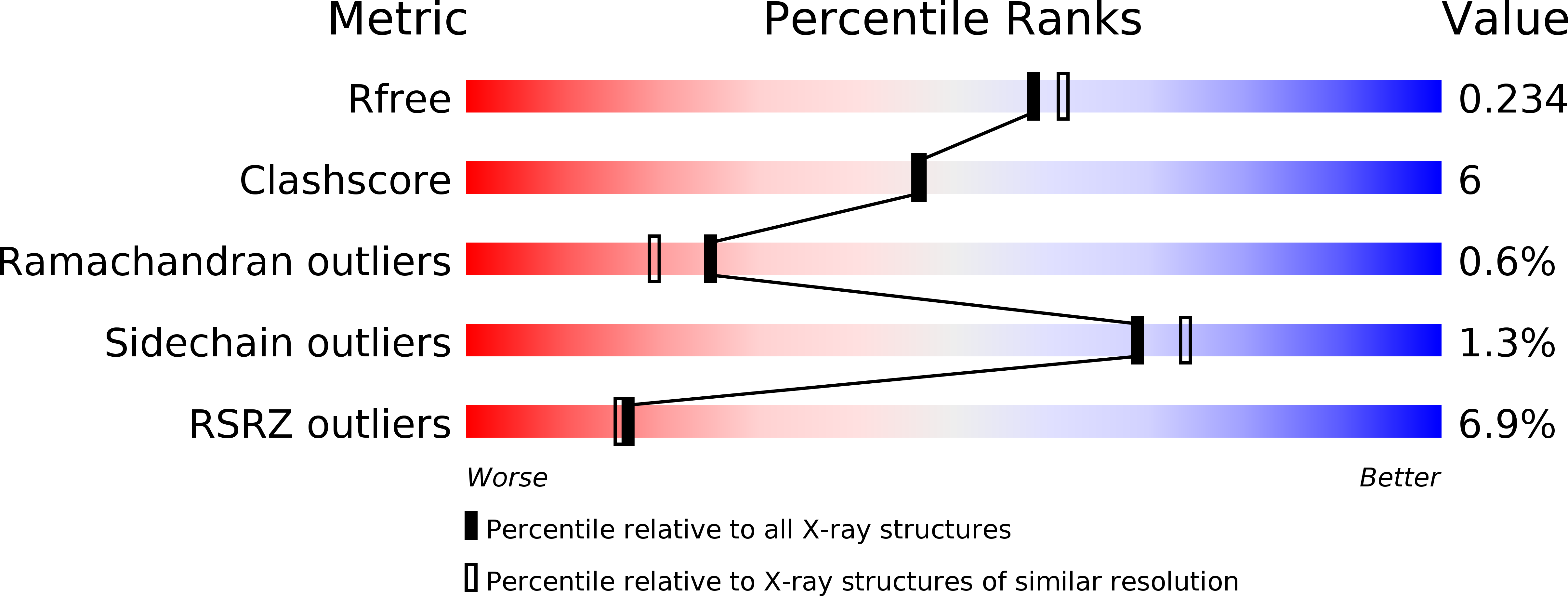
1GL4 image
Deposition Date 2001-08-23
Release Date 2001-11-28
Last Version Date 2024-10-23
Entry Detail
PDB ID:
1GL4
Title:
Nidogen-1 G2/Perlecan IG3 Complex
Biological Source:
Source Organism:
MUS MUSCULUS (Taxon ID: 10090)
Host Organism:
Method Details:
Experimental Method:
Resolution:
2.00 Å
R-Value Free:
0.24
R-Value Work:
0.21
R-Value Observed:
0.21
Space Group:
P 21 21 21
Macromolecular Entities
Structures with similar UniProt ID
Protein Blast
Polymer Type:polypeptide(L)
Molecule:NIDOGEN-1
Gene (Uniprot):Nid1
Chain IDs:A
Chain Length:285
Number of Molecules:1
Biological Source:MUS MUSCULUS
Structures with similar UniProt ID
Protein Blast
Polymer Type:polypeptide(L)
Molecule:BASEMENT MEMBRANE-SPECIFIC HEPARAN SULFATE PROTEOGLYCAN CORE PROTEIN
Gene (Uniprot):Hspg2
Chain IDs:B
Chain Length:98
Number of Molecules:1
Biological Source:MUS MUSCULUS
Primary Citation
Structural Basis for the High-Affinity Interaction of Nidogen-1 with Immunoglobulin-Like Domain 3 of Perlecan
Embo J. 20 5342 ? (2001)
PMID: 11574465 DOI: 10.1093/EMBOJ/20.19.5342

Abstact

Nidogen and perlecan are large multifunctional basement membrane (BM) proteins conserved in all metazoa. Their high-affinity interaction, which is likely to contribute to BM assembly and function, is mediated by the central G2 domain in nidogen and the third immunoglobulin (IG)-like domain in perlecan, IG3. We have solved the crystal structure at 2.0 A resolution of the mouse nidogen-1 G2-perlecan IG3 complex. Perlecan IG3 belongs to the I-set of the IG superfamily and binds to the wall of the nidogen-1 G2 beta-barrel using beta-strands C, D and F. Nidogen-1 residues participating in the extensive interface are highly conserved, whereas the corresponding binding site on perlecan is more variable. We hypothesize that a second, as yet unidentified, activity of nidogen overlaps with perlecan binding and accounts for the unusually high degree of surface conservation in the G2 domain.

Legend

Protein

Chemical

Disease

Primary Citation of related structures