Deposition Date
1996-07-16
Release Date
1997-01-27
Last Version Date
2024-10-30
Entry Detail
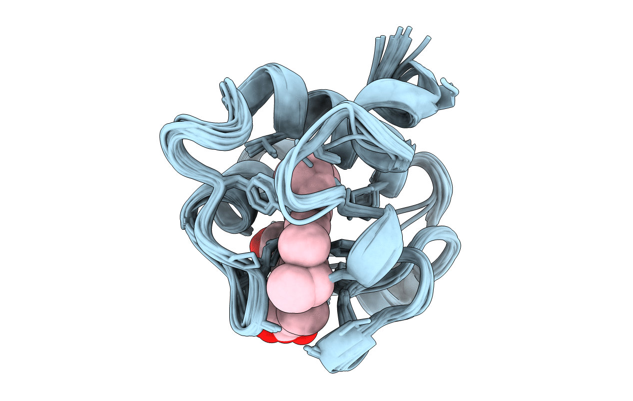
PDB ID:
1GKS
Keywords:
Title:
ECTOTHIORHODOSPIRA HALOPHILA CYTOCHROME C551 (REDUCED), NMR, 37 STRUCTURES
Biological Source:
Source Organism(s):
Halorhodospira halophila (Taxon ID: 1053)
Method Details:
Experimental Method:
Conformers Calculated:
40
Conformers Submitted:
37
Selection Criteria:
PHYSICAL AND EXPERIMENTAL ENERGY