Deposition Date
2001-08-20
Release Date
2002-06-27
Last Version Date
2025-04-09
Entry Detail
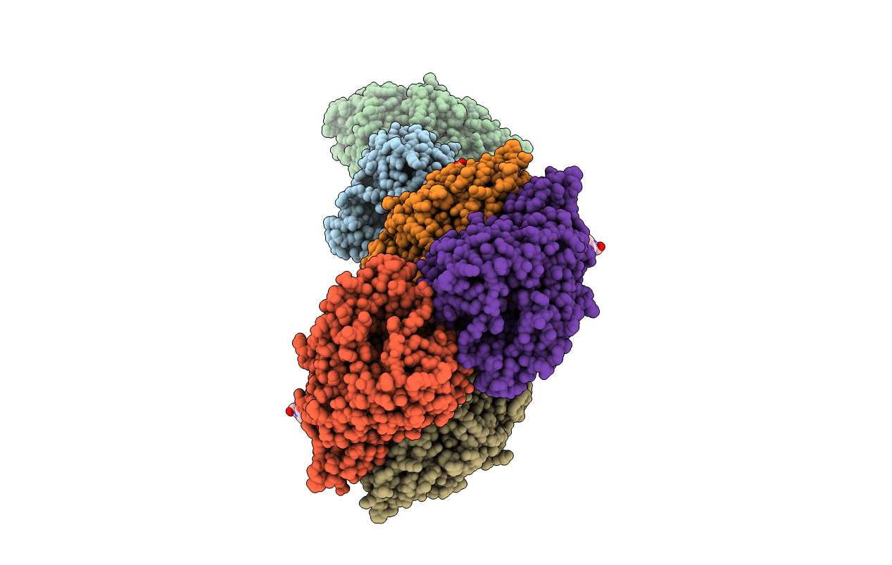
PDB ID:
1GKP
Keywords:
Title:
D-Hydantoinase (Dihydropyrimidinase) from Thermus sp. in space group C2221
Biological Source:
Source Organism(s):
THERMUS SP. (Taxon ID: 275)
Expression System(s):
Method Details:
Experimental Method:
Resolution:
1.30 Å
R-Value Free:
0.18
R-Value Work:
0.15
R-Value Observed:
0.15
Space Group:
C 2 2 21