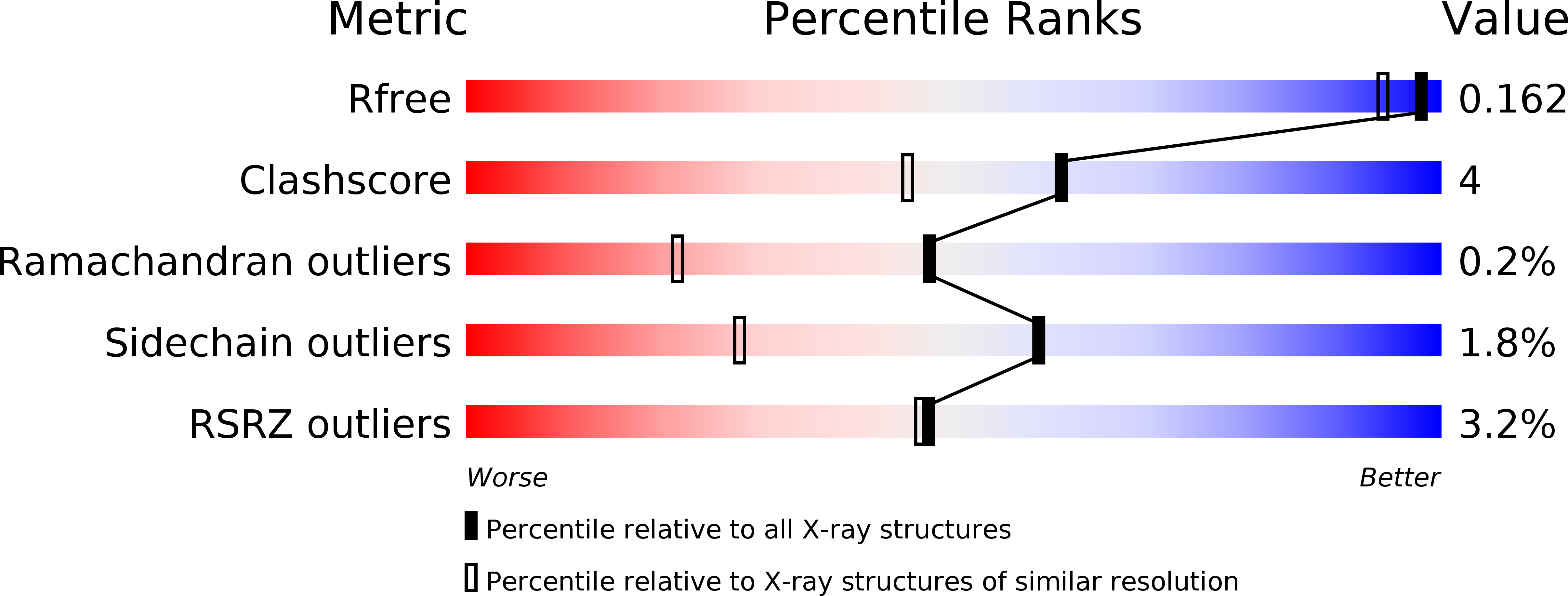
1GK8 image
Deposition Date 2001-08-09
Release Date 2001-10-24
Last Version Date 2023-12-13
Entry Detail
PDB ID:
1GK8
Keywords:
Title:
Rubisco from Chlamydomonas reinhardtii
Biological Source:
Source Organism:
Method Details:
Experimental Method:
Resolution:
1.40 Å
R-Value Free:
0.16
R-Value Work:
0.14
R-Value Observed:
0.14
Space Group:
C 1 2 1
Macromolecular Entities
Polymer Type:polypeptide(L)
Molecule:RIBULOSE-1,5 BISPHOSPHATE CARBOXYLASE LARGE CHAIN
Gene (Uniprot):rbcL
Chain IDs:A, B (auth: C), C (auth: E), D (auth: G)
Chain Length:475
Number of Molecules:4
Biological Source:CHLAMYDOMONAS REINHARDTII
Polymer Type:polypeptide(L)
Molecule:RIBULOSE BISPHOSPHATE CARBOXYLASE SMALL CHAIN 1
Gene (Uniprot):RBCS1
Chain IDs:E (auth: I), F (auth: K), G (auth: M), H (auth: O)
Chain Length:140
Number of Molecules:4
Biological Source:CHLAMYDOMONAS REINHARDTII
Modified Residue
Compound ID Chain ID Parent Comp ID Details 2D Image
HYP A PRO 4-HYDROXYPROLINE
KCX A LYS LYSINE NZ-CARBOXYLIC ACID
MME E MET N-METHYL METHIONINE
SMC A CYS S-METHYLCYSTEINE
Primary Citation
First Crystal Structure of Rubisco from a Green Alga, Chlamydomonas Reinhardtii.
J.Biol.Chem. 276 48159 ? (2001)
PMID: 11641402 DOI: 10.1074/JBC.M107765200

Abstact

The crystal structure of Rubisco (ribulose 1,5-bisphosphate carboxylase/oxygenase) from the unicellular green alga Chlamydomonas reinhardtii has been determined to 1.4 A resolution. Overall, the structure shows high similarity to the previously determined structures of L8S8 Rubisco enzymes. The largest difference is found in the loop between beta strands A and B of the small subunit (betaA-betaB loop), which is longer by six amino acid residues than the corresponding region in Rubisco from Spinacia. Mutations of residues in the betaA-betaB loop have been shown to affect holoenzyme stability and catalytic properties. The information contained in the Chlamydomonas structure enables a more reliable analysis of the effect of these mutations. No electron density was observed for the last 13 residues of the small subunit, which are assumed to be disordered in the crystal. Because of the high resolution of the data, some posttranslational modifications are unambiguously apparent in the structure. These include cysteine and N-terminal methylations and proline 4-hydroxylations.

Legend

Protein

Chemical

Disease

Primary Citation of related structures