1GJQ image
Deposition Date 2001-08-01
Release Date 2002-08-01
Last Version Date 2024-11-20
Entry Detail
PDB ID:
1GJQ
Keywords:
Title:
Pseudomonas aeruginosa cd1 nitrite reductase reduced cyanide complex
Biological Source:
Source Organism:
Method Details:
Experimental Method:
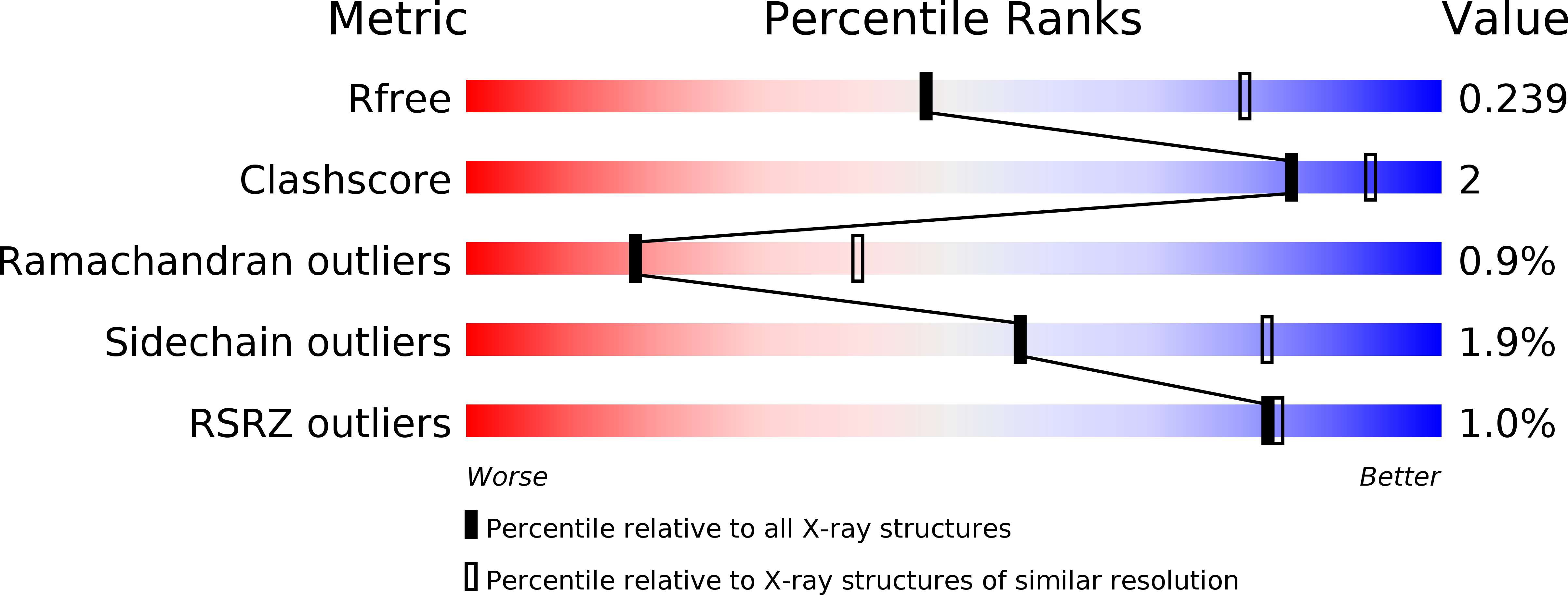
Resolution:
2.70 Å
R-Value Free:
0.24
R-Value Work:
0.20
R-Value Observed:
0.20
Space Group:
P 21 21 2
Macromolecular Entities
Structures with similar UniProt ID
Protein Blast
Polymer Type:polypeptide(L)
Molecule:NITRITE REDUCTASE
Gene (Uniprot):nirS
Chain IDs:A, B
Chain Length:543
Number of Molecules:2
Biological Source:PSEUDOMONAS AERUGINOSA
Primary Citation
Cyanide Binding to Cd(1) Nitrite Reductase from Pseudomonas Aeruginosa: Role of the Active-Site His369 in Ligand Stabilization.
Biochem.Biophys.Res.Commun. 291 1 ? (2002)
PMID: 11829453 DOI: 10.1006/BBRC.2002.6391

Abstact

Cyanide binding to fully reduced Pseudomonas aeruginosa cd(1) nitrite reductase (Pa cd(1) NiR) has been investigated for the wild-type enzyme and a site-directed mutant in which the active-site His369 was replaced by Ala. This mutation reduces the affinity toward cyanide (by approximately 13-fold) and especially decreases the rate of binding of cyanide to the reduced d(1) heme (by approximately 100-fold). The crystal structure of wild-type reduced Pa cd(1) NiR saturated with cyanide was determined to a resolution of 2.7 A. Cyanide binds to the iron of the d(1) heme, with an Fe-C-N angle of 168 degrees for both subunits of the dimer and only His369 is within hydrogen bonding distance of the nitrogen atom of the ligand. These results suggest that in Pa cd(1) NiR the invariant distal residue His369 plays a dominant role in controlling the binding of anionic ligands and allow the discussion of the mechanism of cyanide binding to the wild-type enzyme.

Legend

Protein

Chemical

Disease

Primary Citation of related structures
Feedback Form
Name
Email
Institute
Feedback