1GIO image
Deposition Date 1996-04-12
Release Date 1996-12-07
Last Version Date 2024-10-30
Entry Detail
PDB ID:
1GIO
Title:
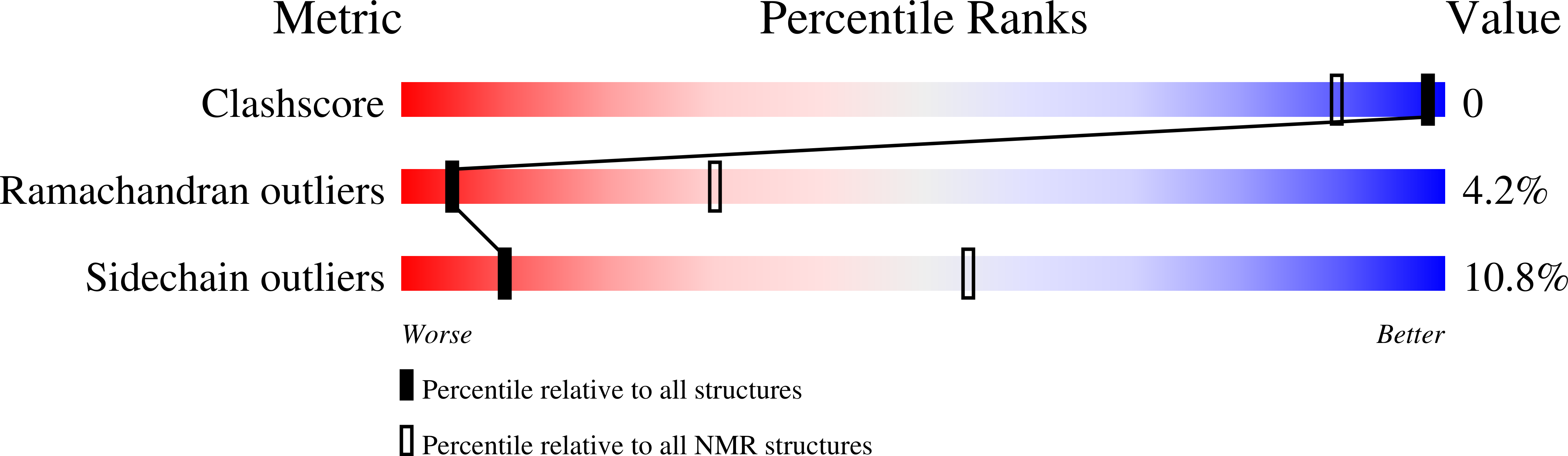
NMR SOLUTION STRUCTURE OF BOVINE ANGIOGENIN, 10 STRUCTURES
Biological Source:
Source Organism:
Bos taurus (Taxon ID: 9913)
Method Details:
Experimental Method:
Conformers Submitted:
10
Macromolecular Entities
Structures with similar UniProt ID
Protein Blast
Polymer Type:polypeptide(L)
Molecule:ANGIOGENIN
Gene (Uniprot):ANG1
Chain IDs:A
Chain Length:125
Number of Molecules:1
Biological Source:Bos taurus
Ligand Molecules
Primary Citation
Solution structure of bovine angiogenin by 1H nuclear magnetic resonance spectroscopy.
Biochemistry 35 8870 8880 (1996)
PMID: 8688423 DOI: 10.1021/bi960022r

Abstact

The three-dimensional structure of bovine angiogenin has been determined using two- and three-dimensional proton NMR spectroscopy. The solution structure is very close to that recently determined by X-ray diffraction analysis. This structure appears well defined, even if five loops and one helix exhibit greater flexibility. Analysis of the active site geometry confirms the position of the Glu-118 residue which obstructs the pyrimidine binding site. There is no experimental evidence of an unobstructed conformation of angiogenin in solution. In addition, it appears that the Glu-118 and Ser-119 residues and the cell receptor binding loop may play an important role in the differences of C-terminal fragment organization and ribonucleolytic activity observed between angiogenins and ribonuclease A.

Legend

Protein

Chemical

Disease

Primary Citation of related structures