1GH4 image
Deposition Date 2000-11-09
Release Date 2001-05-09
Last Version Date 2024-11-20
Entry Detail
PDB ID:
1GH4
Keywords:
Title:
Structure of the triple mutant (K56M, K120M, K121M) of phospholipase A2
Biological Source:
Source Organism:
Bos taurus (Taxon ID: 9913)
Host Organism:
Method Details:
Experimental Method:
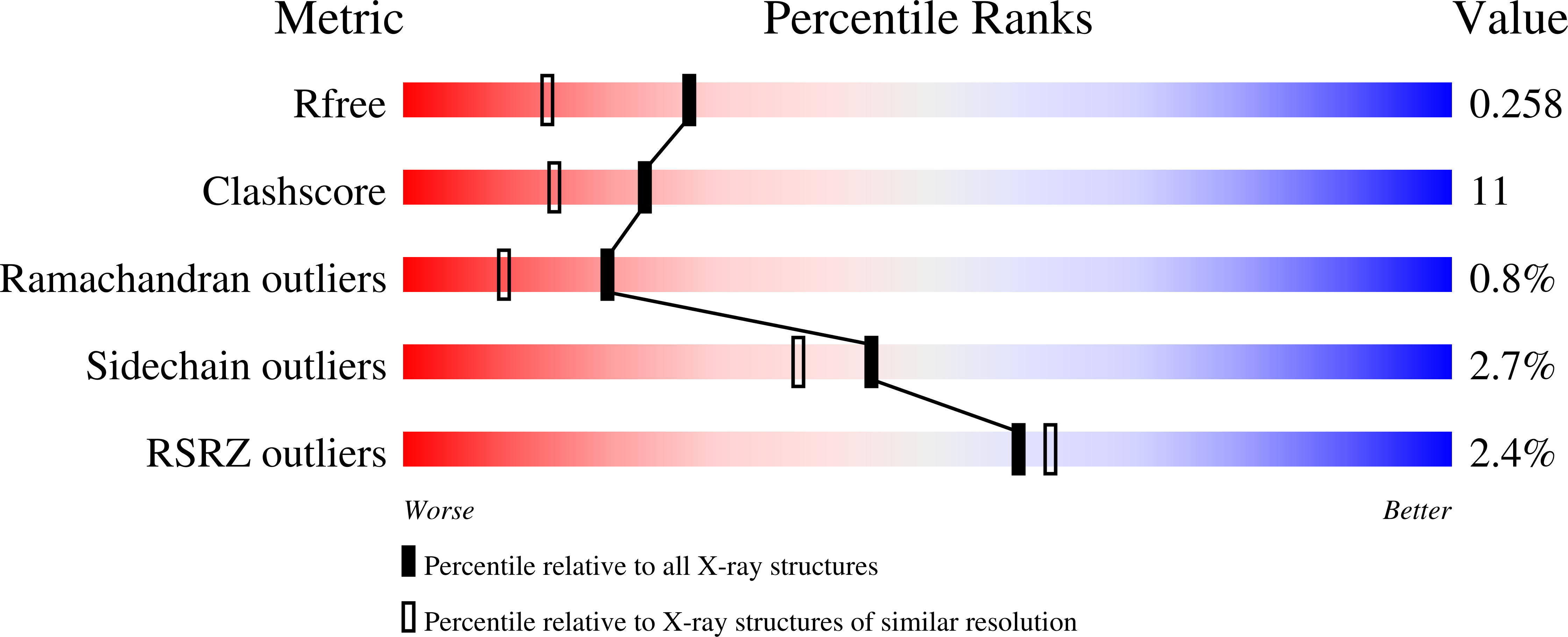
Resolution:
1.90 Å
R-Value Free:
0.25
R-Value Work:
0.19
Space Group:
P 1 2 1
Macromolecular Entities
Structures with similar UniProt ID
Protein Blast
Polymer Type:polypeptide(L)
Molecule:PHOSPHOLIPASE A2
Gene (Uniprot):PLA2G1B
Mutagens:K56M,K120M,K121M
Chain IDs:A
Chain Length:123
Number of Molecules:1
Biological Source:Bos taurus
Primary Citation
Observation of additional calcium ion in the crystal structure of the triple mutant K56,120,121M of bovine pancreatic phospholipase A2.
J.Mol.Biol. 324 755 762 (2002)
PMID: 12460575 DOI: 10.1016/S0022-2836(02)01132-4

Abstact

Phospholipase A(2) catalyses hydrolysis of the ester bond at the C2 position of 3-sn-phosphoglycerides. Here we report the 1.9A resolution crystal structure of the triple mutant K56,120,121M of bovine pancreatic phospholipase A(2). The structure was solved by molecular replacement method using the orthorhombic form of the recombinant phospholipase A(2). The final protein model contains all the 123 amino acid residues, two calcium ions, 125 water molecules and one 2-methyl-2-4-pentanediol molecule. The model has been refined to a crystallographic R-factor of 19.6% (R(free) of 25.9%) for all data between 14.2A and 1.9A. The residues 62-66, which are in a surface loop, are always disordered in the structures of bovine pancreatic phospholipase A(2) and its mutants. It is interesting to note that the residues 62-66 in the present structure is ordered and the conformation varies substantially from those in the previously published structures of this enzyme. An unexpected and interesting observation in the present structure is that, in addition to the functionally important calcium ion in the active site, one more calcium ion is found near the N terminus. Detailed structural analyses suggest that binding of the second calcium ion could be responsible for the conformational change and the ordering of the surface loop. Furthermore, the results suggest a structural reciprocity between the k(cat)(*) allosteric site and surface loop at the i-face, which represents a newly identified structural property of secreted phospholipase A(2).

Legend

Protein

Chemical

Disease

Primary Citation of related structures