Deposition Date
2000-11-10
Release Date
2001-02-28
Last Version Date
2023-12-27
Entry Detail
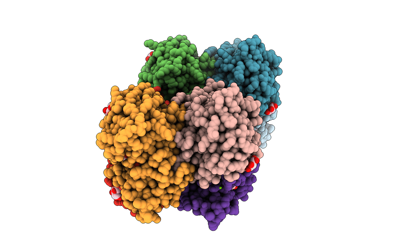
PDB ID:
1GEG
Keywords:
Title:
CRYATAL STRUCTURE ANALYSIS OF MESO-2,3-BUTANEDIOL DEHYDROGENASE
Biological Source:
Source Organism(s):
Klebsiella pneumoniae (Taxon ID: 573)
Expression System(s):
Method Details:
Experimental Method:
Resolution:
1.70 Å
R-Value Free:
0.20
R-Value Work:
0.19
Space Group:
P 1 21 1