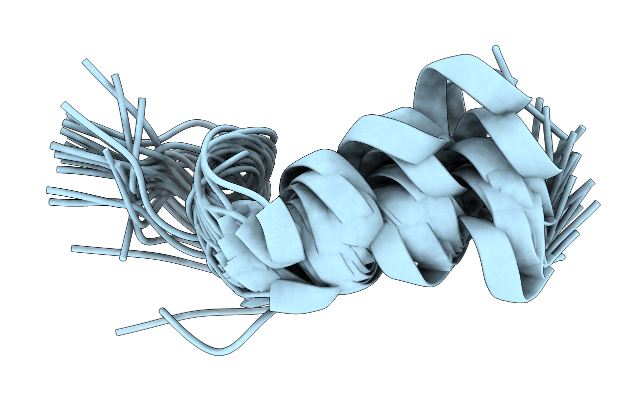
1GEA image
Deposition Date 2000-10-20
Release Date 2001-04-20
Last Version Date 2023-12-27
Entry Detail
PDB ID:
1GEA
Keywords:
Title:
RECEPTOR-BOUND CONFORMATION OF PACAP21
Biological Source:
Source Organism:
(Taxon ID: )
Method Details:
Experimental Method:
Conformers Calculated:
200
Conformers Submitted:
25
Selection Criteria:
structures with the lowest energy
Macromolecular Entities
Polymer Type:polypeptide(L)
Molecule:PITUITARY ADENYLATE CYCLASE ACTIVATING POLYPEPTIDE
Gene (Uniprot):ADCYAP1
Chain IDs:A
Chain Length:21
Number of Molecules:1
Biological Source:
Modified Residue
Compound ID Chain ID Parent Comp ID Details 2D Image
LYN A LYS 2,6-DIAMINO-HEXANOIC ACID AMIDE
Ligand Molecules
Primary Citation
Conformation of a peptide ligand bound to its G-protein coupled receptor.
Nat.Struct.Biol. 8 161 165 (2001)
PMID: 11175907 DOI: 10.1038/84159

Abstact

Many peptide hormones elicit a wide array of physiological effects by binding to G-protein coupled receptors. We have determined the conformation of pituitary adenylate cyclase activating polypeptide, PACAP(1--21)NH(2), bound to a PACAP-specific receptor by NMR spectroscopy. Residues 3--7 form a unique beta-coil structure that is preceded by an N-terminal extended tail. This beta-coil creates a patch of hydrophobic residues that is important for receptor binding. In contrast, the C-terminal region (residues 8--21) forms an alpha-helix, similar to that in the micelle-bound PACAP. Thus, the conformational difference between PACAP in the receptor-bound and the micelle-bound states is limited to the N-terminal seven residues. This observation is consistent with the two-step ligand transportation model in which PACAP first binds to the membrane nonspecifically and then diffuses two-dimensionally in search of its receptor; a conformational change at the N-terminal region then allows specific interactions between the ligand and the receptor.

Legend

Protein

Chemical

Disease

Primary Citation of related structures