Deposition Date
1994-09-14
Release Date
1995-02-27
Last Version Date
2024-02-07
Entry Detail
PDB ID:
1GDL
Keywords:
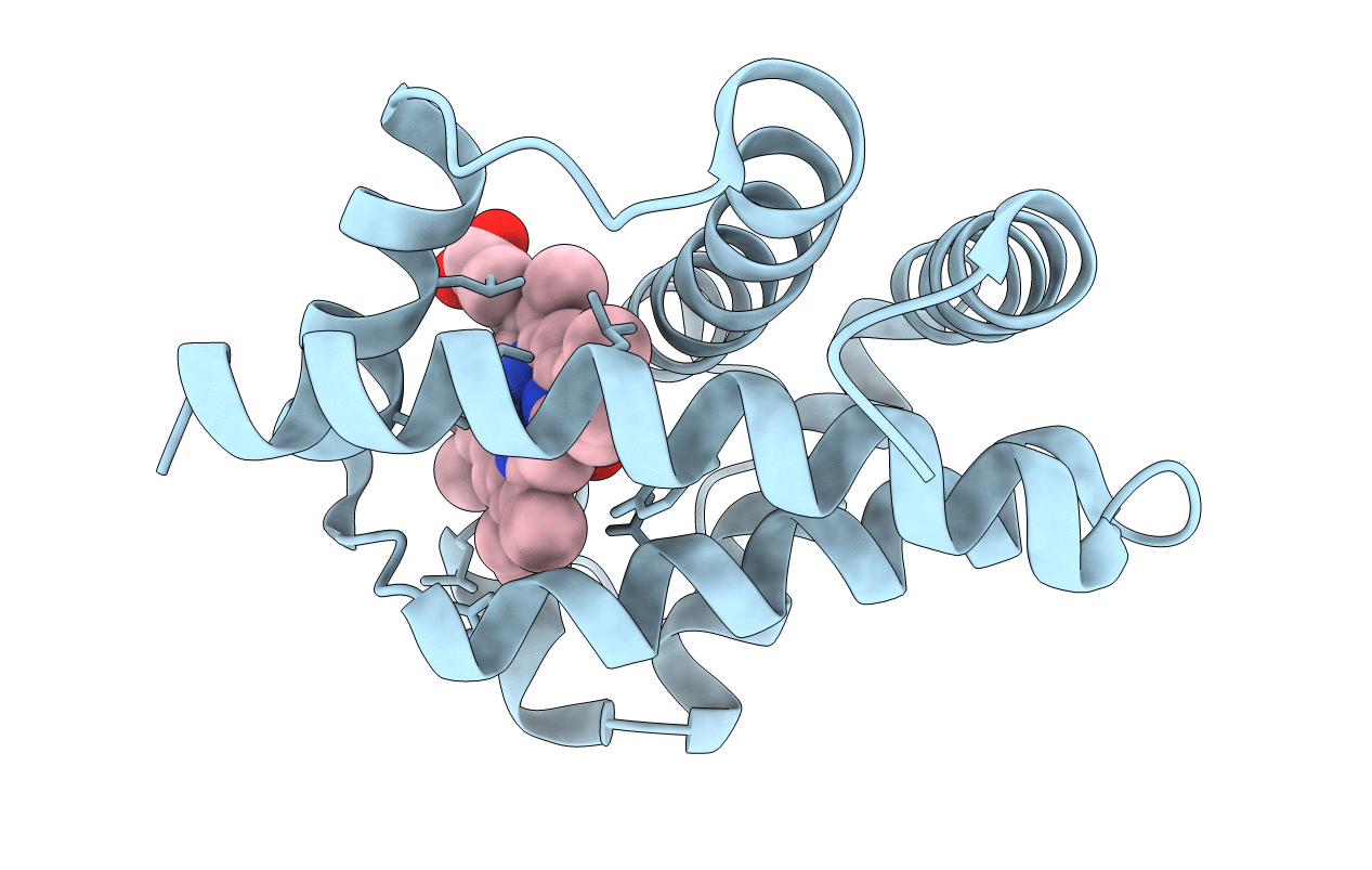
Title:
CRYSTAL STRUCTURE OF FERRIC COMPLEXES OF THE YELLOW LUPIN LEGHEMOGLOBIN WITH ISOQUINOLINE AT 1.8 ANGSTROMS RESOLUTION (RUSSIAN)
Biological Source:
Source Organism(s):
Lupinus luteus (Taxon ID: 3873)
Method Details:
Experimental Method:
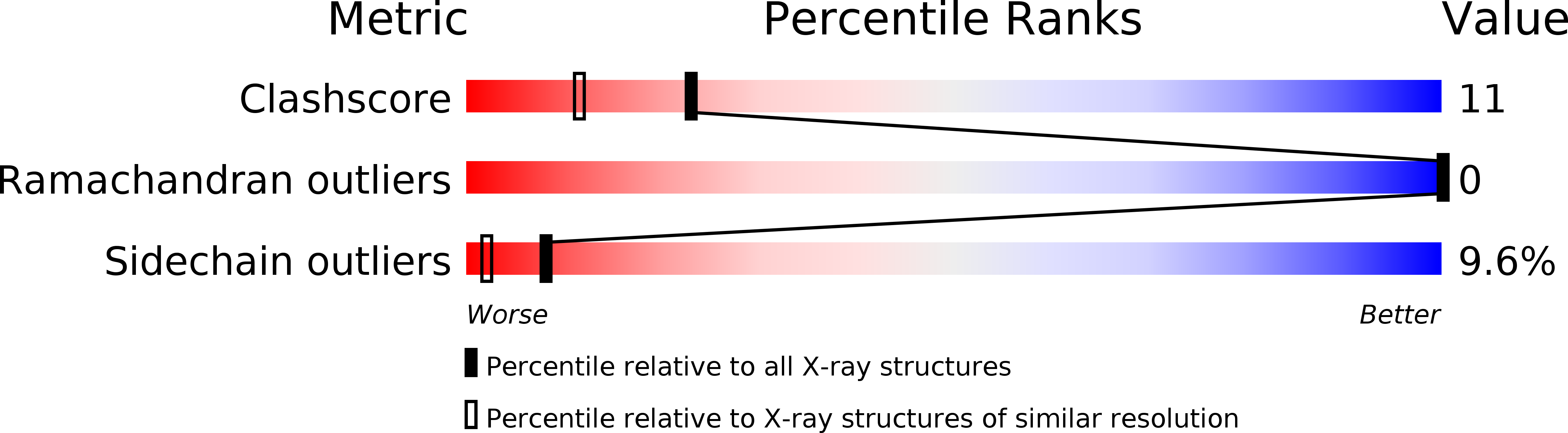
Resolution:
1.80 Å
R-Value Observed:
0.17
Space Group:
C 1 2 1