Deposition Date
2000-08-07
Release Date
2001-02-28
Last Version Date
2023-12-27
Entry Detail
PDB ID:
1GCO
Keywords:
Title:
CRYSTAL STRUCTURE OF GLUCOSE DEHYDROGENASE COMPLEXED WITH NAD+
Biological Source:
Source Organism(s):
Bacillus megaterium (Taxon ID: 1404)
Expression System(s):
Method Details:
Experimental Method:
Resolution:
1.70 Å
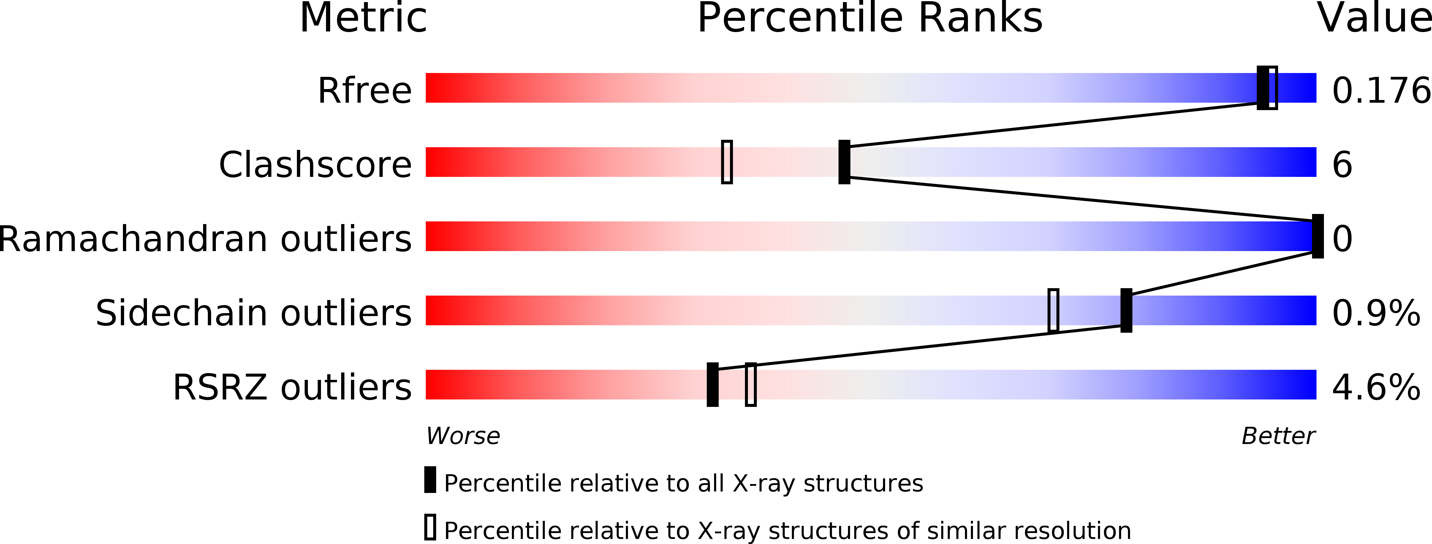
R-Value Free:
0.19
R-Value Work:
0.17
R-Value Observed:
0.17
Space Group:
C 1 2 1