Abstact
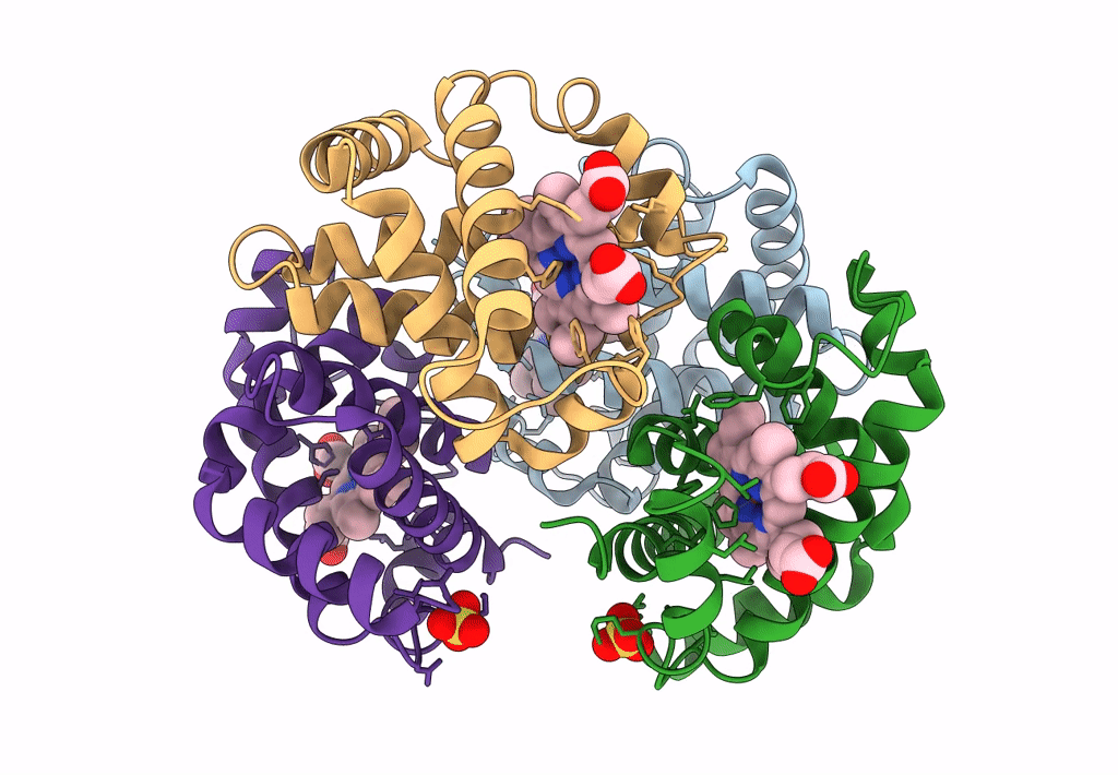
Three variants of tetrameric human hemoglobin, with changes at the alpha1beta2/alpha2beta1-interface, at the alpha1beta1/alpha2beta2-interface, and at both interfaces, have been constructed. At alpha1beta2/alpha2beta1-interface the beta93 cysteine was replaced by alanine (betaC93A), and at the alpha1beta1/alpha2beta2-interface the beta112 cysteine was replaced by glycine (betaC112G). The alpha1beta2 interface variant, betaC93A, and the alpha1beta1/alpha1beta2 double mutant, beta(C93A+C112G), were crystallized in the T-state, and the structures determined at 2. 0 and 1.8 A resolution, respectively. A comparison of the structures with that of natural hemoglobin A shows the absence of detectable changes in the tertiary folding of the protein or in the T-state quaternary assembly. At the beta112 site, the void left by the removal of the cysteine side chain is filled by a water molecule, and the functional characteristics of betaC112G are essentially those of human hemoglobin A. At the beta93 site, water molecules do not replace the cysteine side chain, and the alanine substitution increases the conformational freedom of beta146His, weakening the important interaction of this residue with beta94Asp. As a result, when Cl- is present in the solution, at a concentration 100 mM, the Bohr effect of the two mutants carrying the beta93Cys-->Ala substitution, betaC93A and beta(C93A+C112G), is significantly modified being practically absent below pH 7.4. Based on the crystallographic data, we attribute these effects to the competition between beta94Asp and Cl- in the salt link with beta146His in T-state hemoglobin. These results point to an interplay between the betaHis146-betaAsp94 salt bridge and the Cl- in solution regulated by the Cys present at position beta93, indicating yet another role of beta93 Cys in the regulation of hemoglobin function.