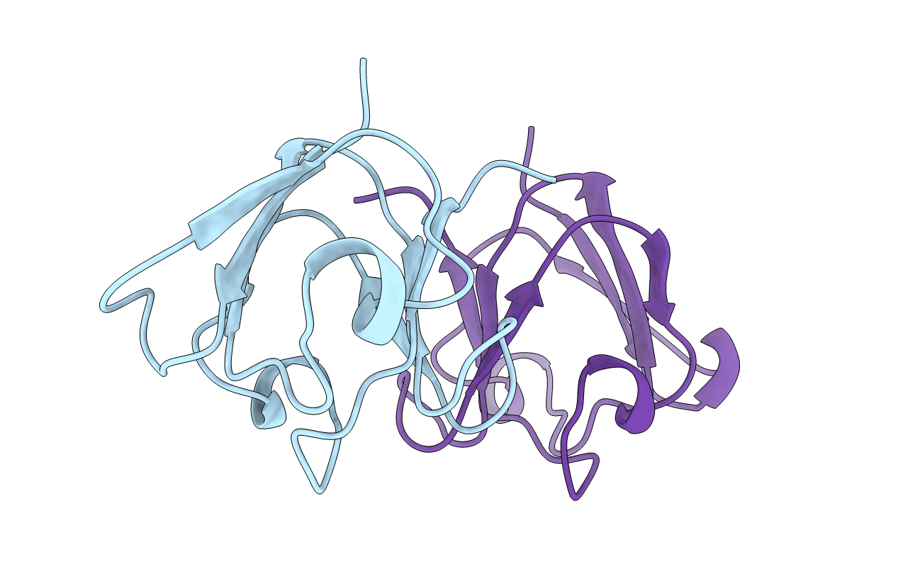
1GAM image
Deposition Date 1996-02-02
Release Date 1996-07-11
Last Version Date 2024-02-07
Entry Detail
PDB ID:
1GAM
Title:
GAMMA B CRYSTALLIN TRUNCATED C-TERMINAL DOMAIN
Biological Source:
Source Organism:
Bos taurus (Taxon ID: 9913)
Host Organism:
Method Details:
Experimental Method:
Resolution:
2.60 Å
R-Value Work:
0.21
R-Value Observed:
0.21
Space Group:
P 32 2 1
Macromolecular Entities
Structures with similar UniProt ID
Protein Blast
Polymer Type:polypeptide(L)
Molecule:GAMMA B CRYSTALLIN
Gene (Uniprot):CRYGB
Chain IDs:A, B
Chain Length:86
Number of Molecules:2
Biological Source:Bos taurus
Primary Citation
The X-ray structures of two mutant crystallin domains shed light on the evolution of multi-domain proteins.
Nat.Struct.Biol. 3 267 274 (1996)
PMID: 8605629 DOI: 10.1038/nsb0396-267

Abstact

We use protein engineering and crystallography to simulate aspects of the early evolution of beta gamma-crystallins by observing how a single domain oligomerizes in response to changes in a sequence extension. The crystal structure of the C-terminal domain of gamma beta-crystallin with its four-residue C-terminal extension shows that the domain does not form a symmetric homodimer analogous to the two-domain pairing in beta gamma-crystallins. Instead the C-terminal extension now forms heterologous interactions with other domains leading to the solvent exposure of the natural hydrophobic interface with a consequent loss in protein solubility. However, this domain truncated by just the C-terminal tyrosine forms a symmetric homodimer of domains in the crystal lattice.

Legend

Protein

Chemical

Disease

Primary Citation of related structures