Deposition Date
1996-03-06
Release Date
1996-08-17
Last Version Date
2024-10-30
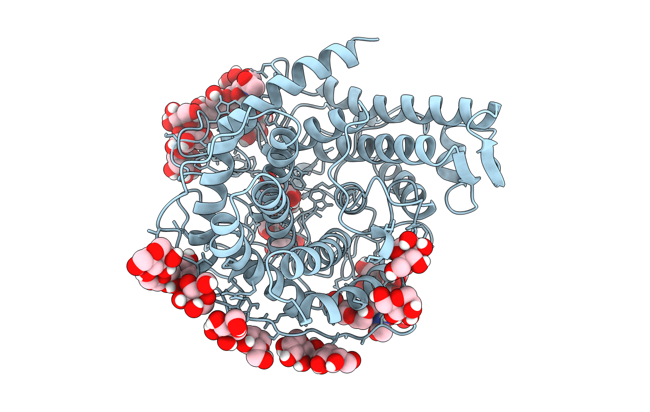
Method Details:
Experimental Method:
Resolution:
2.00 Å
R-Value Free:
0.15
R-Value Work:
0.13
R-Value Observed:
0.13
Space Group:
P 21 21 21