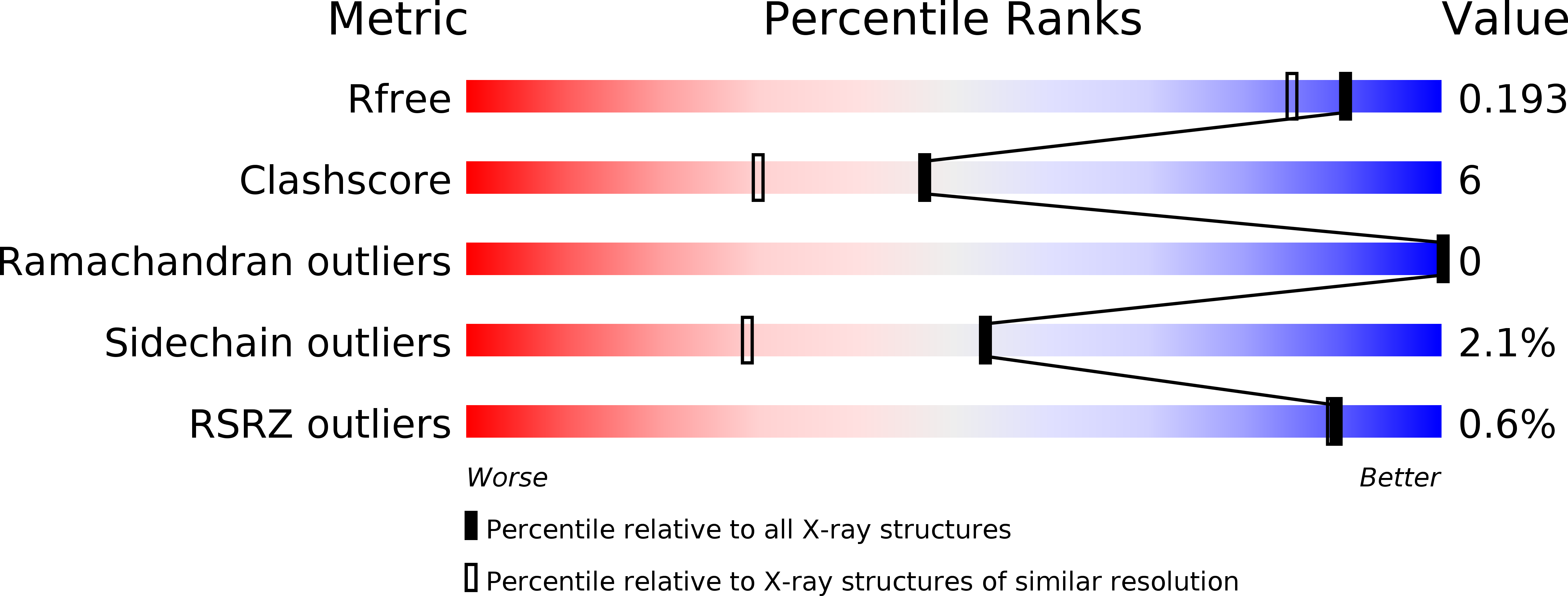
1GA0 image
Deposition Date 2000-11-28
Release Date 2001-06-06
Last Version Date 2024-11-13
Entry Detail
PDB ID:
1GA0
Keywords:
Title:
STRUCTURE OF THE E. CLOACAE GC1 BETA-LACTAMASE WITH A CEPHALOSPORIN SULFONE INHIBITOR
Biological Source:
Source Organism:
Host Organism:
Method Details:
Experimental Method:
Resolution:
1.60 Å
R-Value Free:
0.20
R-Value Work:
0.18
R-Value Observed:
0.18
Space Group:
P 21 21 2
Macromolecular Entities
Structures with similar UniProt ID
Protein Blast
Polymer Type:polypeptide(L)
Molecule:BETA-LACTAMASE
Chain IDs:A
Chain Length:364
Number of Molecules:1
Biological Source:Enterobacter cloacae
Primary Citation
Inhibition of class C beta-lactamases: structure of a reaction intermediate with a cephem sulfone.
Biochemistry 40 6233 6239 (2001)
PMID: 11371184 DOI: 10.1021/bi010131s

Abstact

The crystallographic structure of the Enterobacter cloacae GC1 extended-spectrum class C beta-lactamase, inhibited by a new 7-alkylidenecephalosporin sulfone, has been determined by X-ray diffraction at 100 K to a resolution of 1.6 A. The crystal structure was solved by molecular replacement using the unliganded structure [Crichlow et al. (1999) Biochemistry 38, 10256-10261] and refined to a crystallographic R-factor equal to 0.183 (R(free) 0.208). Cryoquenching of the reaction of the sulfone with the enzyme produced an intermediate that is covalently bound via Ser64. After acylation of the beta-lactam ring, the dihydrothiazine dioxide ring opened with departure of the sulfinate. Nucleophilic attack of a side chain pyridine nitrogen atom on the C6 atom of the resultant imine yielded a bicyclic aromatic system which helps to stabilize the acyl enzyme to hydrolysis. A structural assist to this resonance stabilization is the positioning of the anionic sulfinate group between the probable catalytic base (Tyr150) and the acyl ester bond so as to block the approach of a potentially deacylating water molecule. Comparison of the liganded and unliganded protein structures showed that a major movement (up to 7 A) and refolding of part of the Omega-loop (215-224) accompanies the binding of the inhibitor. This conformational flexibility in the Omega-loop may form the basis of an extended-spectrum activity of class C beta-lactamases against modern cephalosporins.

Legend

Protein

Chemical

Disease

Primary Citation of related structures